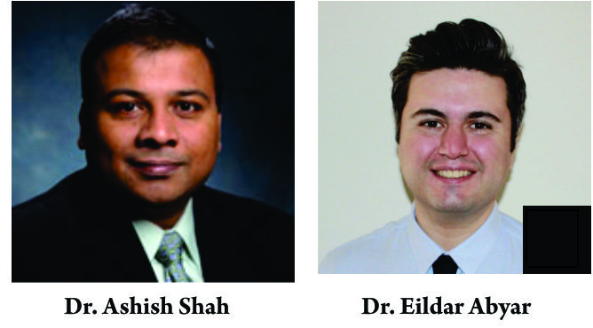
Eildar Abyar, Ashish Shah
Volume 3 | Issue 2 | May – Aug 2018 | Page 30-37
Author: Eildar Abyar [1], Ashish Shah [1].
[1] Department of Orthopaedic Surgery, University of Alabama at Birmingham
Address of Correspondence
Dr. Eildar Abyar,
Department of Orthopaedic Surgery, University of Alabama at Birmingham, USA.
Email: elder.abiar@gmail.com
Abstract
Arthroscopy of the foot and ankle has become an important therapeutic tool for the management of foot and ankle pathologies. Advantages of the arthroscopic technique over open techniques include low post-operative morbidity and absence of limb-threatening complications, less blood loss, shorter hospital stay, faster rehabilitation and mobilization, and a decreased complication rate. To achieve these advantages the surgeon should be thoroughly skilled and familiar with the anatomy of the region3 and arthroscopic techniques. Arthroscopic surgery and tendoscopy are emerging procedures for management of several disorders of the ankle and subtalar joint. These techniques can be both diagnostic and therapeutic and preserve the soft-tissue envelope to a much greater extent than open surgery. The purpose of this review article is to survey the literature regarding the adjunct use of arthroscopy in the treatment of foot and ankle pathologies with highlights in ankle arthroscopy indications and techniques.
Keywords: Foot, Ankle, Arthroscopy,
References
1. O’Brien TS, Hart TS, Shereff MJ, Stone J, Johnson J. Open versus arthroscopic ankle arthrodesis: a comparative study. Foot Ankle Int 1999 Jun;20(6):368-74.
2. Willits K, Sonneveld H, Amendola A, Giffin JR, Griffin S, Fowler PJ. Outcome of posterior ankle arthroscopy for hindfoot impingement. Arthroscopy 2008 Feb;24(2):196-202.
3. Barber FA, Click J, Britt BT. Complications of ankle arthroscopy. Foot Ankle 1990 Apr;10(5):263-6.
4. Watanabe M. Selfoc Arthroscope (Watanabe no 24 Arthroscopes). Tokyo, Japan: Teishin Hospital; 1972.
5. Ferkel RD, Orwin JF. Ankle arthroscopy: a new tool for treating acute and chronic ankle fractures (abtract). Arthroscopy. 1993;9:352-353.
6. Van Dijk CN, Verhagen RA, Tole JL. Arthroscopy for problems after ankle fracture. J Bone Joint Surg Br. 1997;79: 280-284.
7. Ferkel RD, Small HN, Gittins JE. Complications in foot and ankle arthroscopy. Clin Orthop Relat Res 2001 Oct;(391):89-
104.
8. Van Dijk CN, Scholte D. Arthroscopy of the ankle joint.
Arthroscopy 1997 Feb;13(1):90-6.
9. Van Dijk CN, Scholten PE, Krips R. A 2-portal endoscopic approach for diagnosis and treatment of posterior ankle pathol- ogy. Arthroscopy 2000 Nov;16(8):871-6.
10. Stutz G, Kuster MS, Kleinstück F, Gächter A: Arthroscopic management of septic arthritis: stages of infection and results, Knee Surg Sports Traumatol Arthrosc 8:270–274, 2000.
11. Waguri-Nagaya Y, Kobayashi M, Goto H, et al: Septic arthritis of the right ankle caused by Staphylococcus aureus infection in a rheumatoid arthritis patient treated with etanercept, Mod Rheumatol 17:338–0, 2007.
12. Kleiger B. Anterior tibiotalar impingement syndromes in dancers. Foot Ankle 3:69–73, 1982.
13. van den Bekerom MP, Raven EE. The distal fascicle of the anterior inferior tibiofibular ligament as a cause of tibiotalar impingement syndrome: a current concepts review. Knee Surg Sports Traumatol Arthrosc 15:465–471, 2007.
14. Umans H. Ankle impingement syndromes. Semin Musculoskelet Radiol 6:133–139, 2002.
15. Renstrom PAFH. Persistently painful sprained ankle. J Am Acad Orthop Surg 2:270–280, 1994.
16. Morris LH. Athlete’s ankle. J Bone Joint Surg 25:220–223, 1943.
17. McMurray TP. Footballer’s ankle. J Bone Joint Surg 32[Br]:68–69, 1950.
18. Ferkel RD, Karzel RP, Del Pizzo W, Friedman MJ, Fischer SP. Arthroscopic treatment of anterolateral impingement of the ankle. Am J Sports Med 19:440–446, 1991.
19. Deberardino TM, Arciero RA, Taylor DC. Arthroscopic treatment of soft tissue impingement of the ankle in athletes. Arthroscopy 13:492–498, 1997.
20. Wolin I, Glassman F, Sideman F, Levinthal DH. Internal derangement of talofibular component of the ankle. Surg Gynecol Obstet 91:193–200, 1950.
21. Bassett FH, Gates HS, Billys JB, Morris HB, Nikolaou PK. Talar impingement by the anteroinferior tibiofibular ligament. A cause of chronic pain in the ankle after inversion sprain. J Bone Joint Surg 72[Am]:55–59, 1990.
22. Akseki D, P´ynar H, Bozkurt M, Yald´yz K, Araç S. The distal fascicle of the anterior inferior tibiofibular ligament as a cause of anterolateral ankle impingement. Acta Orthop Scand 70:478–482, 1999.
23. Meislin RJ, Rose DJ, Parisien JS, Springer S. Arthroscopic treatment of synovial impingement of the ankle. Am J Sports Med 21:186–189, 1993.
24. Liu SH, Raskin A, Osti L, Baber C, Jacobson K, Finerman G. Arthroscopic treatment of anterolateral ankle impingement. Arthroscopy 10:215–218, 1994.
25. Martin DF, Curl WW, Baker CL. Arthroscopic treatment of chronic synovitis of the ankle. Arthroscopy 5:110–114, 1989.
26. Kim SH, Ha KI. Arthroscopic treatment for impingement of the anterolateral soft tissues of the ankle. J Bone Joint Surg 82[Br]:1019–1021, 2000.
27. Amandola A, Petrik J, Webster-Bogaert S. Ankle arthroscopy: outcome in 79 consecutive patients. Arthroscopy 12:565–573, 1996.
28. Ferkel RD, Karzel RP, Del Pizzo W, et al. Arthroscopic treatment of anterolateral impingement of the ankle. Am J Sports Med. 1991;19(5):440–6.
29. DeBerardino TM, Arciero RA, Taylor DC. Arthroscopic treatment of soft-tissue impingement of the ankle in athletes. Arthroscopy. 1997;13(4):492–8.
30. Kim SH, Ha KI. Arthroscopic treatment for impingement of the anterolateral soft tissues of the ankle. J Bone Joint Surg Br. 2000;82(7):1019–21.
31. Arnold H. Posttraumatic impingement syndrome of the ankle: indications and results of arthroscopic therapy. Foot Ankle Surg. 2011;17:85–8.
32. Tol JL, van Dijk CN. Anterior ankle impingement. Foot Ankle Clin 2006 Jun;11(2):297-310, vi.
33. Ogilvie-Harris DJ, Mahomed N, Demaziere A. Anterior impingement of the ankle treated by arthroscopic removal of bony spurs. J Bone Joint Surg Br. 1993;75:437–40. Seventeen patients who performed arthroscopic resection for anterior impingement were reviewed. Pain, swelling, and limping were improved in a 3 year follow-up.
34. Ferkel RD, Karzel RP, Del Pizzo W, et al. Arthroscopic treatment of anterolateral impingement of the ankle. Am J Sports Med. 1991;19(5):440–6.
35. Biedert R. Anterior ankle pain in sports medicine: aetiology and indications for arthroscopy. Arch Orthop Trauma Surg. 1991;110 (6):293–7.
36. Amendola A, Petrik J, Webster-Bogaert S. Ankle arthroscopy: outcome in 79 consecutive patients. Arthroscopy. 1996;12(5):565–73. Study with seventy nine patients who performed ankle arthroscopic debridement after impingement syndrome with 80-86 % improve after surgery.
37. Scranton PE, McDermott JE. Anterior tibiotalar spurs: a comparison of open versus arthroscopic debridement. Foot Ankle. 1992;13 (3):125–9.
38. Niek van Dijk C: Anterior and posterior ankle impingement. Foot Ankle Clin 2006;11(3):663-683.
39. Scholten PE, Sierevelt IN, van Dijk CN. Hindfoot endoscopy for posterior ankle impingement. J Bone Joint Surg Am 2008 Dec;90(12):2665-72.
40. Willits K, Sonneveld H, Amendola A, Giffin JR, Griffin S, Fowler PJ. Outcome of posterior ankle arthroscopy for hindfoot impingement. Arthroscopy 2008 Feb;24(2):196-202.
41. Hamilton WG, Geppert MJ, Thompson FM. Pain in the posterior aspect of the ankle in dancers. Differential diagnosis and operative treatment. J Bone Joint Surg Am 1996 Oct;78(10):1491-500.
42. Van Dijk CN, Lim LS, Poortman A, Strubbe EH, Marti RK. Degenerative joint disease in female ballet dancers. Am J Sports Med 1995 May;23(3):295-300.
43. Hedrick MR, McBryde AM. Posterior ankle impingement. Foot Ankle Int 1994 Jan;15(1):2-8.
44. Weinstein SL, Bonfiglio M. Unusual accessory (bipartite) talus simulating fracture. A case report. J Bone Joint Surg Am 1975 Dec;57(8):1161-3.
45. Sarrafian SK. Anatomy of the foot and ankle: descriptive, topographic, functional. Philadelphia: Lippincott; 1983.
46. Bizarro AH. On sesamoid and supernumerary bones of the limbs. J Anat 1921;55:256-68.
47. Lapidus PW. A note on the fracture of os trigonum syndrome. Report of a case. Bull Hosp Jt Dis 1972;33(2):150-4.
48. Ferkel RD. Soft-tissue lesions of the ankle. In: Whipple TL, editor. Arthroscopic surgery: the foot and ankle. Philadelphia: Lippincott-Raven; 1996. p. 121-43.
49. SooHoo NF, Zingmond DS, Ko CY. Comparison of reoperation rates following ankle arthrodesis and total ankle arthroplasty. J Bone Joint Surg Am. 2007;89:2143–9.
50. Haddad SL, Coetzee JC, Estok R, et al. Intermediate and long-term outcomes of total ankle arthroplasty and ankle arthrodesis. A systematic review of the literature. J Bone Joint Surg Am. 2007;89:1899–905.
51. Schneider D. Arthroscopic ankle fusion. Arthroscopic Video J. 1983, 3.
52. Zvijac JE, Lemak L, Schurhoff MR, et al. Analysis of arthroscopically assisted ankle arthrodesis. Arthroscopy. 2002;18(1):70–5.
53. Myerson MS, Quill G. Ankle arthrodesis. A comparison of an arthroscopic and an open method of treatment. Clin Orthop Relat Res. 1991;268:84–95.
54. Peterson KS, Lee MS, Buddecke DE. Arthroscopic versus open ankle Arthroscopic versus open ankle arthrodesis: a retrospective cost analysis. J Foot Ankle Surg. 2010;49(3):242–7.
55. Winson IG, Robinson DE, Allen PE. Arthroscopic ankle arthrodesis. J Bone Joint Surg Br. 2005;87-B(3):343–7.
56. Gougoulias NE, Agathangelidis FG, Parsons SW. Arthroscopic ankle arthrodesis. Foot Ankle Int. 2007;28(6):695–706.
57. Dannawi Z, Nawabi DH, Patel A, et al. Arthroscopic ankle arthrodesis: are results reproducible irrespective of pre-operative deformity? Foot Ankle Surg. 2011;17(4):294–9. This paper supports the use arthroscopic tibiotalar fusion in cases of high angular deformity of the ankle.
58. Pierre A, Hulet C, Locker B, et al. Arthroscopic tibio-talar arthrodesis: limitations and indications in 20 patients. Rev Chir Orthop Reparatrice Appar Mot. 2003;89(2):144–51.
59. Ferkel RD, Hewitt M. Long term results of arthroscopic ankle arthrodesis. Foot Ankle Int. 2005;26(4):275–80.
60. Collman DR, Kaas MH, Schuberth JM. Arthroscopic ankle arthrodesis: factors influencing union in 39 consecutive patients. Foot Ankle Int. 2006;27(12):1079–85.
61. McCullough CJ, Venugopal V. Osteochondritis dissecans of the talus: the natural history. Clin Orthop Relat Res. 1979;144:264-268.
62. Scharling M. Osteochondritis dissecans of the talus. Acta Orthop Scand. 1978;49:89-94.
63. Schenck RC Jr, Goodnight JM. Osteochondritis dissecans. J Bone Joint Surg Am. 1996; 78:439-456.
64. Cahill BR. Osteochondritis dissecans of the knee: treatment of juvenile and adult forms. J Am Acad Orthop Surg. 1995; 3:237-247.
65. McCoy AM, Toth F, Dolvik NI, et al. Articular osteochondrosis: a comparison of naturally-occurring human and animal disease. Osteoarthritis Cartilage. 2013;21:1638-47.
66. O’Driscoll SW. The healing and regeneration of articular cartilage. J Bone Joint Surg Am. 1998;80: 1795-1812.
67. Chuckpaiwong B, Berkson EM, Theodore GH. Micro-fracture for osteochondral lesions of the ankle: outcome analysis and outcome predictors of 105 cases. Arthroscopy. 2008; 24:106-112.
68. Kouvalchouk JF, Schneider-Maunoury G, Rodineau J. Osteochondral lesions of the dome of the talus with partial necrosis. Surgical treatment by curettage and filling. Rev Chir Orthop Reparatrice Appar Mot. 1990;76:480-489.
69. Van Dijk CN, Scholten PE, Krips RA. 2-portal endoscopic approach for diagnosis and treatment of posterior ankle pathology. Arthroscopy 2000;16:871-876.
70. Scholten PE, Sierevelt IN, van Dijk CN. Hindfoot endoscopy for posterior ankle impingement. J Bone Joint Surg Am 2008;90:2665-2672.
71. Willits K, Sonneveld H, Amendola A, Giffin JR, Griffin S, Fowler PJ. Outcome of posterior ankle arthroscopy for hindfoot impingement. Arthroscopy 2008;24:196-202.
72. Galla M, Lobenhoffer P. Technique and results of arthroscopic treatment of posterior ankle impingement. Foot Ankle Surg 2011;17:79-84.
73. Tey M, Monllau JC, Centenera JM, Pelfort X. Benefits of arthroscopic tuberculoplasty in posterior ankle impingement syndrome. Knee Surg Sports Traumatol Arthrosc 2007;15:1235-1239.
74. Russell JA, Shave RM, Yoshioka H, Kruse DW, Koutedakis Y, Wyon MA. Magnetic resonance imaging of the ankle in female ballet dancers en pointe. Acta Radiol 2010;6:655-661.
75. Peace KAL, Hillier JC, Hulme A, Healy JC. MRI features of posterior ankle impingement syndrome in ballet dancers: A review of 25 cases. Clin Radiol 2004;59:1025-1033.
76. Russell JA, Kruse DW, Koutedakis Y, McEwan IM, Wyon M. Pathoanatomy of posterior ankle impingement in ballet dancers. Clin Anat 2010;23:613-621.
77. Ferkel RD, Orwin JF. Ankle arthroscopy: a new tool for treating acute and chronic ankle fractures (abtract). Arthroscopy. 1993;9:352-353.
78. Schuberth JM, Cobb MD, Talarico RH. Minimally invasive arthroscopic-assisted reduction with percutaneous fixation in the management of intra-articular calcaneal fractures: a review of 24 cases. J Foot Ankle Surg. 2009;48:315-322.
79. Hintermann B, Regazzoni P, Lampert C, Stutz G, Gachter A. Arthroscopic findings in acute fractures of the ankle. J Bone Joint Surg Br. 2000;82:345-351.
80. Thordarson DB, Bains R, Shepherd LE. The role of ankle arthroscopy on the surgical management of ankle fractures. Foot Ankle Int. 2001;22:123-125.
81. Olszewski AD, Jones R, Farrell R, Kaylor K. The effects of dilute epinephrine saline irrigation on the need for tourniquet use in routine arthroscopic knee surgery. Am J Sports Med. 1999;27:354-356.
82. Schuberth JM, Collman DR, Rush SM, Ford LA. Deltoid ligament integrity in lateral malleolar fractures: a comparative analysis of arthroscopic and radiographic assessments. J Foot Ankle Surg. 2004;43: 20-29.
83. Schuberth JM, Jennings MM, Lau AC. Arthroscopy-assisted repair of latent syndesmotic instability of the ankle. Arthroscopy. 2008;24:868-874.
84. Bonasia DE, Rossi R, Saltzman CL, Amendola A. The role of arthroscopy in the management of fractures about the ankle. J Am Acad Orthop Surg. 2011;19:226-235.
85. Loren GJ, Ferkel RD. Arthroscopic assessment of occult intra-articular injury in acute ankle fractures. Arthroscopy. 2002;18:412-421.
86. Taga I, Shino K, Inoue M, Nakata K, Maeda A. Articular cartilage lesions in ankles with lateral ligament injury. Am J Sports Med. 1993;21:120-127.
87. Aktas S, Kocaoglu B, Gereli A, Nalbantodlu U, Guven O. Incidence of chondral lesions of talar dome in ankle fracture types. Foot Ankle Int. 2008;29:287-292.
88. Leontaritis N, Hinojosa L, Panchbhavi VK. Arthroscopically detected intraarticular lesions associated with acute ankle frctures. J Bone Joint Surg Am. 2009;91:333-339.
89. Sorrento DL, Mlodzienski A. Incidence of lateral talar dome lesions in SER IV ankle fractures. J Foot Ankle Surg. 2000;39:354-358.
90. Lloyd J, Elsayad S, Hariharan K, Tanaka H. Revisiting the concept of talar shift in ankle fractures. Foot Ankle Int. 2006;27:793-796.
91. Ramsey PL, Hamilton W. Changes in tibiotalar area of contact caused by lateral talar shift. J Bone Joint Surg Am.1976;58:356-357.
92. Starkweather MP, Collman DR, Schuberth JM. Early protected weightbearing after open reduction internal fixation of ankle fractures. J Foot Ankle Surg. 2012;51:575-578.
93. Gardner MJ, Demetrakopoulos D, Briggs SM, Helfet DL, Lorich DG. The ability of the Lauge-Hansen classification to predict ligament injury and mechanism in ankle fractures: an MRI study. J Orthop Trauma. 2006;20:267-272.
94. Lui TH, Ip K, Chow HT. Comparison of radiologic and arthroscopic diagnoses of distal tibiofibular syndesmosis disruption in acute ankle fracture. Arthroscopy. 2005;21:1370-1374.
95. Miller AN, Barei DP, Iaquinto JM, Ledoux WR, Beingessner DM. Iatrogenic syndesmosis malreduction via clamp and screw placement. J Orthop Trauma. 2013;27:100-106.
96. Xenos JS, Hopkinson WJ, Mulligan ME, Olson EJ, Popovic NA. The tibiofibular syndesmosis: evaluation of the ligamentous structures, methods of fixation, and radiographic assessment. J Bone Joint Surg Am. 1995;77:847-856.
97. Gardner MJ, Demetrakopoulos D, Briggs SM, Helfet DL, Lorich DG. Malreduction of the tibiofibular syndesmosis in ankle fractures. Foot Ankle Int. 2006;27:788-792.
98. Davidovitch RI, Well Y, Karla R, et al. Intraoperative syndesmotic reduction: three-dimensional versus standard fluoroscopic imaging. J Bone Joint Surg Am. 2013;95:1838-1843.
99. Sri-Ram K, Robinson AH. Arthroscopic assessment of the syndesmosis following ankle fracture. Injury. 2005;36:675-678.
100. Chen SH, Wu PH, Lee YS. Long-term results of pilon fractures. Arch Orthop Trauma Surg. 2007;127:55-60.
101. Conroy J, Agarwal M, Giannoudis PV, Matthews SJE. Early internal fixation and soft tissue cover of severe open tibial pilon fractures. Int Orthop. 2003;27:343-347.
102. Decoster TA, Willis MC, Marsh JL, et al. Rank order analysis of tibial plafond fractures: does injury or reduction predict outcome? Foot Ankle Int. 1999;20:44-49.
103. Kalenderer O, Gunes O, Ozcalabi IT, Ozluk S. Clinical results of tibial pilon fractures treated by open reduction and internal fixation. Acta Orthop Traumatol Turc. 2003;37:133-137.
104. Pollak AN, Mccarthy ML, Bess RS. Outcomes after treatment of high-energy tibial plafond fractures. J Bone Joint Surg Am. 2003;85:1893-1900.
105. Salton HL, Rush S, Schuberth JM. Tibial plafond fractures: limited incision reduction with percutaneous fixation. J Foot Ankle Surg. 2007;46:261-269.
106. Gumann G, Hamilton GA. Arthroscopically assisted treatment of ankle injuries. Clin Podiatr Med Surg. 2009;28:523-538.
107. Ono A, Nishikawa S, Nagao A, et al. Arthroscopically assisted treatment of ankle fractures: arthroscopic findings and surgical outcomes. Arthroscopy. 2004;20:627-631.
108. Hammond AW, Crist BD. Arthroscopic management of C3 tibial plafond fractures: a technical guide. J Foot Ankle Surg. 2012;51:382-386.
109. Kim HS, Jahng JS, Kim SS, Chun CH, Han HJ. Treatment of tibial pilon fractures using ring fixators and arthroscopy. Clin Orthop Relat Res. 1997;334:244-250.
110. Poyanli O, Esenkaya I, Ozkut AT, et al. Minimally invasive reduction technique in split depression type tibial pilon fractures. J Foot Ankle Surg. 2012;51: 254-257.
111. Van Dijk CN, Scholten PE, Krips R: A 2-portal endoscopic approach for diagnosis and treatment of posterior ankle pathology, Arthroscopy 16:871–876, 2000.
112. Acevedo JI, Busch MT, Ganey TM, et al: Coaxial portals for posterior ankle arthroscopy: an anatomic study with clinical correlation on 29 patients, Arthroscopy 16:836–842, 2000.
113. Lohrer H, Arentz S: Posterior approach for arthroscopic treatment of posterolateral impingement syndrome of the ankle in a top-level field hockey player, Arthroscopy 20:e15–e21,
2004.
114. Shurnas PS, Coughlin MJ: Arthritic conditions of the foot. In Coughlin MJ, Mann RA, Saltzman CL, editors: Surgery of the foot and ankle, ed 8, Philadelphia, 2007, Mosby, pp 801–856.
115. Guhl JF. Foot and ankle arthroscopy. New York: Slack; 1993.
116. Martin DF, Baker CL, Curl WW, Andrews JR, Robie DB, Haas AF. Operative ankle arthroscopy. Long-term follow-up. Am J Sports Med 1989 Jan;17(1):16-23.
117. Barber FA, Click J, Britt BT. Complications of ankle arthroscopy. Foot Ankle 1990 Apr;10(5):263-6.
118. Ferkel RD, Heath DD, Guhl JF. Neurological complications of ankle arthroscopy. Arthroscopy 1996 Apr;12(2):200-8.
119. Amendola A, Lee KB, Saltzman CL, Suh JS. Technique and early experience with posterior arthroscopic subtalar arthrodesis. Foot Ankle Int 2007 Mar;28(3):298-302.
120. Andrews JR, Previte WJ, Carson WG. Arthroscopy of the ankle: technique and normal anatomy. Foot Ankle 1985 Aug;6(1):29-33.
121. Drez D, Jr., Guhl JF, Gollehon DL. Ankle arthroscopy: technique and indications. Foot Ankle 1981 Nov;2(3):138-43.
122. Ferkel RD, Fasulo GJ. Arthroscopic treatment of ankle injuries. Orthop Clin North Am 1994 Jan;25(1):17-32.
123. Guhl JF. New concepts (distraction) in ankle arthroscopy. Arthroscopy 1988;4(3):160-7.
124. Unger F, Lajtai G, Ramadani F, Aitzetmuller G, Orthner E. [Arthroscopy of the upper ankle joint. A retrospective analysis of complications]. Unfallchirurg 2000 Oct;103(10):858-63.
| How to Cite this article: Abyar E, Shah A. Foot and Ankle Arthroscopy: Updates, Indications and Technique. Asian Journal Arthroscopy. May-Aug 2018;3(2):30-37. |
Endoscopic Plantar Fasciotomy with Gastrocnemius Recession for Chronic Plantar Fasciitis


HA-BMAC: The Surgical Technique, Pearls and Pitfalls
Katarzyna Herman, Vetri Kumar, Dawid Szwedowski, Luca Chierici, Alberto Gobbi
Volume 4 | Issue 1 | Jan – April 2019 | Page 34- 37
Author: Katarzyna Herman[1], Vetri Kumar[1], Dawid Szwedowski[1], Luca Chierici[1], Alberto Gobbi[1]
Orthopaedic Arthroscopic Surgery International (OASI) Bioresearch Foundation, Milan, Italy.
Address of Correspondence
Dr.Alberto Gobbi, MD
Orthopaedic Arthroscopic Surgery International (OASI) Bioresearch Foundation, Via G.A. Amadeo 24, 20133, Milan, Italy.
Email: gobbi@cartilagedoctor.it
Abstract
Articular cartilage is a highly specialized tissue with poor healing potential. Damage to the cartilage following an injury to the joint is prevalent which leads to osteoarthritis. More research is aimed towards tissue regeneration and prevention of degeneration. Efforts to repair and restore the hyaline like cartilage using two-stage procedures such as autologous chondrocyte implantation have led to the development of scaffolds. Bone marrow aspirate concentrate that contains multi potent stem cells which has the potential to differentiate into hyaline like cartilage along with the use of a scaffold is an effective, reliable and single-stage method of cartilage restoration
References
1.Hunter W. On the Structure and Diseases of Articulating Cartilage. Philos Trans RsocLond B Biol Sci 1743; 9:277.
2. Mankin HJ. The response of articular cartilage to mechanical injury. J Bone Joint Surg Am.1982;64(3):460-6.
3. Widuchowski W, Widuchowski J, Trzaska T(2007) Articular cartilage defects: study of 25,124 knee arthroscopies. Knee; 14:177-82
4. Flanigan DC, Harris JD, Trinh TQ, Siston RA, Brophy RH (2010) Prevalence of chondral defects in athletes’ knees: a systematic review. Med Sci Sports Exerc 42:1795–1801
5. Johnson DL, Urban WP Jr, Caborn DN et al (1998) Articular cartilage changes seen with magnetic resonance imaging-detected bone bruises associated with acute anterior cruciate ligament rupture. Am J Sports Med 26:409–414
6. Lohmander LS, Roos H, Dahlberg L et al (1994) Temporal patterns of stromelysin-1, tissue inhibitor, and proteoglycan fragments in human knee joint fluid after injury to the cruciate ligament or meniscus. J Orthop Res 12:21–28
7. Mandelbaum BR, Browne JE, Fu F et al (1998) Articular carti-lage lesions of the knee. Am J Sports Med 26:853–861
8. Kotlarz H, Gunnarsson CL, Fang H, Rizzo JA. Insurer and out-of-pocket costs of osteoarthritis in the US: evidence from national survey data. Arthritis Rheum. 2009 Dec;60(12):3546-53.
9. Takeda H, Nakagawa T, Nakamura K, Engebretsen L. Prevention and management of knee osteoarthritis and knee cartilage injury in sports. Br J Sports Med. 2011 Apr;45(4):304-9. Epub 2011 Feb 25.
10. Marcacci M, Berruto M, Brocchetta D, et al. Articular cartilage engineering with Hyalograft(R) C: 3-year clinical results. Clin OrthopRelat Res 2005;435:96-105.
11. Gobbi A, Kon E, Berruto M, et al. Patellofemoral full-thickness chondral defects treated with second-generation autologous chondrocyte implantation: Results at 5 years’ follow-up. Am J Sports Med 2009;37: 1083-1092
12. Ando W, Tateishi K, Katakai D, et al. In vitro generation of a scaffold-free tissue-engineered construct (TEC) derived from human synovial mesenchymal stem cells: biological and mechanical properties and further chondrogenic potential. Tissue Eng Part A. 2008;14(12): 2041-2049.
13. Caplan AI. Mesenchymal stem cells: cell-based reconstructive ther-apy in orthopedics. Tissue Eng. 2005;11(7-8):1198-1211.
14. Caplan AI. Mesenchymal stem cells: the past, the present, the future. Cartilage. 2010;1(1):6-9.
15. Dimarino AM, Caplan AI, Bonfield TL. Mesenchymal stem cells in tissue repair. Front Immunol. 2013;4:201.
16. Huselstein C, Li Y, He X. Mesenchymal stem cells for carti-lage engineering. Biomed Mater Eng. 2012;22:69-80.
17. Pasquinelli G, Orrico C, Foroni L, Bonafè F, Carboni M, Guarnieri C, et al. Mesenchymal stem cell interaction with a non-woven hyaluronan- based scaffold suitable for tissue repair. J Anat. 2008;213(5):520-30.
18. Lisignoli G, Cristino S, Piacentini A, Zini N, Noël D, Jorgensen C, et al. Chondrogenic differentiation of murine and human mesenchymal stromal cells in a hyaluronic acid scaffold: differences in gene expression and cell morphology. J Biomed Mater Res A. 2006;77(3):497-506.
19. Facchini A, Lisignoli G, Cristino S, Roseti L, De Franceschi L, Marconi E, et al. Human chondrocytes and mesenchymal stem cells grown onto engineered scaffold. Biorheology. 2006;43(3-4):471-80.
20. Nejadnik H, Hui JH, Feng Choong EP, Tai BC, Lee EH. Autologous bone marrow-derived mesenchymal stem cells versus autologous chondrocyte implantation: an observational cohort study. Am J Sports Med. 2010 Jun; 38(6): 1110-6.
21. Gobbi A, Chaurasia S, Karnatzikos G, Nakamura N. Matrix-Induced Autologous Chondrocyte Implantation versus Multipotent Stem Cells for the Treatment of Large Patellofemoral Chondral Lesions: A Nonrandomized Prospective Trial. Cartilage. 2015 Apr;6(2):82-97.
22. Gobbi A, Karnatzikos G, Sankineani SR. One-step surgery with multipotent stem cells for the treatment of large full-thickness chondral defects of the knee. Am J Sports Med. 2014 Mar;42(3):64857.
23. Gobbi A et al. Biologic Arthroplasty for Full-thickness Cartilage Lesions of the Knee: Results at Three Years Follow-up (SS-56) Arthroscopy , Volume 29 , Issue 6 , e27
24. Gobbi A, Karnatzikos G, Scotti C, Mahajan V, Mazzucco L, Grigolo B. One-Step Cartilage Repair with Bone Marrow Aspirate Concentrated Cells and Collagen Matrix in Full-Thickness Knee Cartilage Lesions: Results at 2-Year Follow-up. Cartilage.2011 Jul;2(3):286-99.
25. Gobbi A, Scotti C, Karnatzikos G, Mudhigere A, Castro M, Peretti GM. One-step surgery with multipotent stem cells and Hyaluronan-based scaffold for the treatment of full-thickness chondral defects of the knee in patients older than 45 years. Knee Surgery, Sports Traumatology, Arthroscopy. 2017;25(8):2494-2501.
26. Whyte GP, Gobbi A, Sadlik B. Dry Arthroscopic Single-Stage Cartilage Repair of the Knee Using a Hyaluronic Acid-Based Scaffold With Activated Bone Marrow-Derived Mesenchymal Stem Cells. Arthroscopy Techniques. 2016;5(4):e913-e918.
(Abstract) (Full Text HTML) (Download PDF)
Gel Based Autologous Chondrocyte Implantation: The Surgical Technique
Deepak Goyal, Vishvas Modi
Volume 4 | Issue 1 | Jan – April 2019 | Page 27- 33
Author: Deepak Goyal[1], Vishvas Modi[1]
Saumya Arthroscopy & Sports Knee Clinic,
Ahmedabad, India.
Address of Correspondence
Dr (Prof) Deepak Goyal
Saumya Arthroscopy & Sports Knee Clinic,
Ahmedabad, India.
Email: deepak@knee.in
Abstract
Autologous chondrocyte implantation (ACI) is one such technique that has the ability to provide a hyaline (like) repair of the localized cartilage lesions, even when they are of a big size. However, the procedure must be chosen very wisely because of its stringent indications and contraindications. Decision to do ACI procedure is very crucial and the surgeon must come to the decision after a detailed clinic-radiological examination. Gel based ACI is one such technique that allows a 3-dimensional distribution of the autologous cultured chondrocytes in a scaffold that is made of fibrin glue. The technique takes away the common complications that were associated with 1st and 2nd generation ACI; like graft hypertrophy, poor access to the lesion, membrane suturing, monolayer distribution etc. The purpose of this paper is to discuss the indications, contraindications, decision making and preoperative planning for the gel based autologous chondrocyte implantation technique in detail along with the surgical procedure, postoperative rehabilitation and the possible complications.
References
1. Ossendorf C, Kaps C, Kreuz PC, Burmester GR, Sittinger M, Erggelet C. Treatment of posttraumatic and focal osteoarthritic cartilage defects of the knee with autologous polymer-based three-dimensional chondrocyte grafts: 2-year clinical results. Arthritis Res Ther. 2007;9(2):R41.
2. Oussedik S, Tsitskaris K, Parker D. Treatment of articular cartilage lesions of the knee by microfracture or autologous chondrocyte implantation: a systematic review. Arthroscopy. 2015;31(4):732-744.
3. Goyal D, Keyhani S, Lee EH, Hui JHP. Evidence-Based Status of Microfracture Technique: A Systematic Review of Level I and II Studies. Arthroscopy. 2013;29(9):1579-1588.
4. Bark S, Riepenhof H, Gille J. AMIC Cartilage Repair in a Professional Soccer Player. Case Rep Orthop. 2012;2012:364342.
5. Gille J, Behrens P, Schulz AP, Oheim R, Kienast B. Matrix-Associated Autologous Chondrocyte Implantation: A Clinical Follow-Up at 15 Years. Cartilage. 2016;7(4):309-315.
6. Minas T, Ogura T, Bryant T. Autologous Chondrocyte Implantation. JBJS Essent Surg Tech. 2016;6(2):e24.
7. Bartlett W, Skinner JA, Gooding CR, et al. Autologous chondrocyte implantation versus matrix-induced autologous chondrocyte implantation for osteochondral defects of the knee: a prospective, randomised study. J Bone Joint Surg Br. 2005;87(5):640-645.
8. Behrens P, Bitter T, Kurz B, Russlies M. Matrix-associated autologous chondrocyte transplantation/implantation (MACT/MACI)–5-year follow-up. Knee. 2006;13(3):194-202.
9. Pavesio A, Abatangelo G, Borrione A, et al. Hyaluronan-based scaffolds (Hyalograft C) in the treatment of knee cartilage defects: preliminary clinical findings. Novartis Found Symp. 2003;249:203-217; discussion 229-233, 234-238, 239-241.
10. Schneider U, Rackwitz L, Andereya S, et al. A prospective multicenter study on the outcome of type I collagen hydrogel-based autologous chondrocyte implantation (CaReS) for the repair of articular cartilage defects in the knee. Am J Sports Med. 2011;39(12):2558-2565.
11. Niemeyer P, Albrecht D, Andereya S, et al. Autologous chondrocyte implantation (ACI) for cartilage defects of the knee: A guideline by the working group “Clinical Tissue Regeneration” of the German Society of Orthopaedics and Trauma (DGOU). Knee. 2016;23(3):426-435.
12. Goyal D, Goyal A, Adachi N. Subchondral Bone: Healthy Soil for the Healthy Cartilage. In: Gobbi A, Espregueira-Mendes J, Lane JG, Karahan M, eds. Bio-Orthopaedics. Berlin, Heidelberg: Springer Berlin Heidelberg; 2017:479-486.
13. Cole BJ, Corpus KT, Bajaj S, et al. Prospective Evaluation of Autologous Chondrocyte Implantation Procedure: Minimum Seven-Year Follow-Up (SS-26). Arthroscopy: The Journal of Arthroscopic & Related Surgery. 2011;27(5):e43-e44.
14. Goyal D, Goyal A, Keyhani S, Lee EH, Hui JHP. Evidence-based status of second- and third-generation autologous chondrocyte implantation over first generation: a systematic review of level I and II studies. Arthroscopy. 2013;29(11):1872-1878.
15. Goyal D, Keyhani S, Goyal A, Lee EH, Hui JHP, Vaziri AS. Evidence-Based Status of Osteochondral Cylinder Transfer Techniques: A Systematic Review of Level I and II Studies. Arthroscopy. 2014;30(4):497-505.
16. Pietschmann MF, Horng A, Niethammer T, et al. Cell quality affects clinical outcome after MACI procedure for cartilage injury of the knee. Knee Surg Sports Traumatol Arthrosc. 2009;17(11):1305-1311.
17. Vanlauwe J, Saris DBF, Victor J, et al. Five-year outcome of characterized chondrocyte implantation versus microfracture for symptomatic cartilage defects of the knee: early treatment matters. Am J Sports Med. 2011;39(12):2566-2574.
18. Gillogly SD, Myers TH, Reinold MM. Treatment of full-thickness chondral defects in the knee with autologous chondrocyte implantation. J Orthop Sports Phys Ther. 2006;36(10):751-764.
19. Hamby TS, Gillogly SD, Peterson L. Treatment of patellofemoralarticular cartilage injuries with autologous chondrocyte implantation. Operative Techniques in Sports Medicine. 2002;10(3):129-135.
20. Rosenberg TD, Paulos LE, Parker RD, Coward DB, Scott SM. The forty-five-degree posteroanterior flexion weight-bearing radiograph of the knee. J Bone Joint Surg Am. 1988;70(10):1479-1483.
21. Zbojniewicz AM, Laor T. Imaging of osteochondritis dissecans. Clin Sports Med. 2014;33(2):221-250.
22. Karim AR, Cherian JJ, Jauregui JJ, Pierce T, Mont MA. Osteonecrosis of the knee: review. Ann Transl Med. 2015;3(1):6.
23. Houpt JB, Pritzker KP, Alpert B, Greyson ND, Gross AE. Natural history of spontaneous osteonecrosis of the knee (SONK): a review. Semin Arthritis Rheum. 1983;13(2):212-227.
24. Kellgren JH, Lawrence JS. Radiological assessment of osteo-arthrosis. Ann Rheum Dis. 1957;16(4):494-502.
25. Altman R, Asch E, Bloch D, et al. Development of criteria for the classification and reporting of osteoarthritis. Classification of osteoarthritis of the knee. Diagnostic and Therapeutic Criteria Committee of the American Rheumatism Association. Arthritis Rheum. 1986;29(8):1039-1049.
26. Spector TD, Hart DJ, Byrne J, Harris PA, Dacre JE, Doyle DV. Definition of osteoarthritis of the knee for epidemiological studies. Ann Rheum Dis. 1993;52(11):790-794.
27. Goyal D, Goyal A, Adachi N. Joint Preservation Surgery for Medial Compartment Osteoarthritis. Arthrosc Tech. 2017;6(3):e717-e728.
28. Recht MP, Resnick D. Magnetic resonance imaging of articular cartilage: an overview. Top Magn Reson Imaging. 1998;9(6):328-336.
29. De Smet AA, Fisher DR, Graf BK, Lange RH. Osteochondritis dissecans of the knee: value of MR imaging in determining lesion stability and the presence of articular cartilage defects. AJR Am J Roentgenol. 1990;155(3):549-553.
30. Lecouvet FE, van de Berg BC, Maldague BE, et al. Early irreversible osteonecrosis versus transient lesions of the femoral condyles: prognostic value of subchondral bone and marrow changes on MR imaging. AJR Am J Roentgenol. 1998;170(1):71-77.
31. Mont MA, Baumgarten KM, Rifai A, Bluemke DA, Jones LC, Hungerford DS. Atraumatic osteonecrosis of the knee. J Bone Joint Surg Am. 2000;82(9):1279-1290.
32. Björkengren AG, AlRowaih A, Lindstrand A, Wingstrand H, Thorngren KG, Pettersson H. Spontaneous osteonecrosis of the knee: value of MR imaging in determining prognosis. AJR Am J Roentgenol. 1990;154(2):331-336.
33. Healy WL. Osteonecrosis of the knee detected only by magnetic resonance imaging. Orthopedics. 1991;14(6):703-704.
34. Goyal D, Goyal A, Brittberg M. Consideration of religious sentiments while selecting a biological product for knee arthroscopy. Knee Surg Sports Traumatol Arthrosc. 2013;21(7):1577-1586.
35. Goyal D, Palkhiwala B. Cover Image. Arthroscopy: The Journal of Arthroscopic & Related Surgery. 2013;29(3):A9.
36. Goyal D. The overlay ACI technique for extra-large osteochondral lesions of the knee joint. 2nd Congress of Asian Cartilage Repair Society at Seoul, Korea, 2014.
37. Choi N-Y, Kim B-W, Yeo W-J, et al. Gel-type autologous chondrocyte (Chondron) implantation for treatment of articular cartilage defects of the knee. BMC Musculoskelet Disord. 2010;11:103.
38. Gillogly SD, Voight M, Blackburn T. Treatment of articular cartilage defects of the knee with autologous chondrocyte implantation. J Orthop Sports Phys Ther. 1998;28(4):241-251.
39. Reinold MM, Wilk KE, Dugas JR, Cain EL, Gillogly SD. Rehabilitation Guidelines: Autologous Chondrocyte Implantation Using Carticel. Cambridge, MA: Genzyme Biosurgery; 2004.
40. Reinold MM, Wilk KE, Macrina LC, Dugas JR, Cain EL. Current concepts in the rehabilitation following articular cartilage repair procedures in the knee. J Orthop Sports Phys Ther. 2006;36(10):774-794.
41. Peterson L, Minas T, Brittberg M, Nilsson A, Sjögren-Jansson E, Lindahl A. Two- to 9-year outcome after autologous chondrocyte transplantation of the knee. Clin Orthop Relat Res. 2000;(374):212-234.
42. Peterson L, Brittberg M, Kiviranta I, Akerlund EL, Lindahl A. Autologous chondrocyte transplantation. Biomechanics and long-term durability. Am J Sports Med. 2002;30(1):2-12.
43. Peterson L, Vasiliadis HS, Brittberg M, Lindahl A. Autologous chondrocyte implantation: a long-term follow-up. Am J Sports Med. 2010;38(6):1117-1124.
44. Pareek A, Carey JL, Reardon PJ, Peterson L, Stuart MJ, Krych AJ. Long-Term Outcomes after Autologous Chondrocyte Implantation: A Systematic Review at Mean Follow-Up of 11.4 Years. Cartilage. 2016;7(4):298-308.
45. Kim MK, Choi SW, Kim SR, Oh IS, Won MH. Autologous chondrocyte implantation in the knee using fibrin. Knee Surg Sports Traumatol Arthrosc. 2010;18(4):528-534.
46. Wood JJ, Malek MA, Frassica FJ, et al. Autologous cultured chondrocytes: adverse events reported to the United States Food and Drug Administration. J Bone Joint Surg Am. 2006;88(3):503-507.
47. Niemeyer P, Pestka JM, Kreuz PC, et al. Characteristic complications after autologous chondrocyte implantation for cartilage defects of the knee joint. Am J Sports Med. 2008;36(11):2091-2099.
(Abstract) (Full Text HTML) (Download PDF)
Scaffold based Autologous Chondrocyte Implantation: The Surgical Technique
Professor Mats Brittberg
Volume 4 | Issue 1 | Jan – April 2019 | Page 23- 26
Author: Professor Mats Brittberg[1]
[1] Cartilage Research Unit, University of Gothenburg, Region Halland Orthopaedics, Kungsbacka Hospital, S-434 80 Kungsbacka Sweden.
Address of Correspondence
Professor- Mats Brittberg
Cartilage Research Unit, University of Gothenburg, Region Halland Orthopaedics, Kungsbacka Hospital, S-434 80 Kungsbacka Sweden.
Email: Mats.brittberg@telia.com
Abstract
Autologous chondrocyte implantation (ACI) has been used clinically since 1987. Most reports on ACI are on the first and second generation ACI with cells in suspension under a sutured membrane and performed with open surgery. Today’s 3rd and 4th generation ACI with cells seeded and grown in or on scaffolds prior to implantation opens up for transarthroscopic implantations. Transarthroscopic surgery reduces the morbidity for the patients and also fastens up the rehab process. In this paper, techniques used for scaffold-based ACI are presented.
References
1.Brittberg M, Lindahl A, Nilsson A et al.Treatment of deep cartilage defects in the knee with autologous chondrocyte transplantation. New England Journal of Medicine 1994; 331:889-895.
2.Brittberg M.Cartilage repair. On cartilaginous tissue engineering with the emphasis on chondrocyte transplantation. Thesis. University of Gothenburg.1996.
3.Brittberg M. Cell carriers as the next generation of cell therapy for cartilage repair: a review of the matrix-induced autologous chondrocyte implantation procedure.Am J Sports Med. 2010; Jun;38(6):1259-71.
4.Steinwachs M. New technique for cell-seeded collagen-matrix-supported autologous chondrocyte transplantation. Arthroscopy. 2009; Feb;25(2):208-11.
5.Hendriks JA, Moroni L, Riesle et al. The effect of scaffold-cell entrapment capacity and physico-chemical properties on cartilage regeneration. Biomaterials. 2013 Jun;34(17):4259-65
6.de Windt TS, Vonk LA, Slaper-Cortenbach ICM, Nizak R, van Rijen MHP, Saris DBF. Allogeneic MSCs and Recycled Autologous Chondrons Mixed in a One-Stage Cartilage Cell Transplantion: A First-in-Man Trial in 35 PatientsStem Cells. 2017 Aug;35(8):1984-1993
7.Brittberg M. Cartilage fragment implantation. . In Cartilage Surgery. An operative manual. Eds Brittberg M, Gersoff W. Elsevier Saunders Philadelphia,2011; 181-191.
8.Brittberg M, Concaro S. Transarthroscopic implantation of Hyalograft (Hyaff 11) with autologous chondrocytes. In Cartilage Surgery. An operative manual. Eds Brittberg M, Gersoff W. Elsevier Saunders Philadelphia,2011; 147-160.
9. Nehrer S, Dorotka R, Domayer S et al. Treatment of full-thickness chondral defects with hyalograft C in the knee: a prospective clinical case series with 2 to 7 years’ follow-up. Am J Sports Med. 2009; Nov;37 Suppl 1:81S-87S
10.Vascellari A, Rebuzzi E, Schiavetti et al. Implantation of matrix-induced autologous chondrocyte (MACI(®)) grafts using carbon dioxide insufflation arthroscopy. Knee Surg Sports Traumatol Arthrosc. 2014 Jan;22(1):219-25.
11.Marcacci M, Berruto M, Brocchetta D et al. Articular cartilage engineering with Hyalograft C: 3-year clinical results. Clin Orthop Relat Res.2005; Jun;(435):96-105
12.Della Villa S, Kon E, Filardo G et al. Does intensive rehabilitation permit early return to sport without compromising the clinical outcome after arthroscopic autologous chondrocyte implantation in highly competitive athletes? Am J Sports Med. 2010; Jan;38(1):68-77.
(Abstract) (Full Text HTML) (Download PDF)
The Mosaicplasty / OAT procedure: Technique, Pearls and Pitfalls
Rajkumar Amaravathi, Renato Andrade, Ricardo Bastos, João Espregueira-Mendes
Volume 4 | Issue 1 | Jan – April 2019 | Page 15-22
Author: Rajkumar Amaravathi [1], Renato Andrade [2,3,4], Ricardo Bastos [2,3,5,6], João Espregueira-Mendes [2]
[1] Department Of Orthopedics, Division of Arthroscopy and Sports Surgery, St. John’s Medical College and Hospital, Bangalore 560034,India
[2] Clínica do Dragão, Espregueira-Mendes Sports Centre – FIFA Medical Centre of Excellence, Porto, Portugal.
[3] Dom Henrique Research Centre, Porto, Portugal.
[4] Faculty of Sports, University of Porto, Porto, Portugal.
[5] Fluminense Federal University, Niteroi, Brazil.
[6] ICVS/3B’s-PT Government Associate Laboratory, Guimarães, Portugal.
[7] School of Medicine, Minho University, Braga, Portugal.
Address of Correspondence
Dr João Espregueira-Mendes,
M.D., Ph.D.; Clínica do Dragão, Espregueira-Mendes Sports Centre – FIFA Medical Centre of Excellence,
Estádio do Dragão, Entrada Nascente, Piso -3, 4350-415, Porto, Portugal.
Email: espregueira@dhresearchcentre.com
Abstract
Osteochondral autologous transplantation is a surgical procedure that involves the transplant of the autologous cartilage from the non-weight bearing areas of the knee to the articular defect. It has the advantage of being a single stage procedure, repairs the subchondral bone, provides hyaline cartilage and allows a fast return to play. It is indicated for small and medium-sized defects, but the mosaicplasty technique allows treating defects up to 9 cm2. A major disadvantage of this technique is the donor site morbidity associated with the graft harvesting. To overcome this drawback, we harvest the autografts from the upper tibio-fibular joint with low or none donor site morbidity. Osteochondral autologous transplantation and mosaicplasty procedures remain an excellent option for small to medium osteochondral injuries resulting in long-term good to excellent clinical and imaging outcomes.Osteochondral autologous transplantation is a surgical procedure that involves the transplant of the autologous cartilage from the non-weight bearing areas of the knee to the articular defect. It has the advantage of being a single stage procedure, repairs the subchondral bone, provides hyaline cartilage and allows a fast return to play. It is indicated for small and medium-sized defects, but the mosaicplasty technique allows treating defects up to 9 cm2. A major disadvantage of this technique is the donor site morbidity associated with graft harvesting. To overcome this drawback, we harvest the autografts from the upper tibio-fibular joint with low or none donor site morbidity. Osteochondral autologous transplantation and mosaicplasty procedures remain an excellent option for small to medium osteochondral injuries resulting in long-term good to excellent clinical and imaging outcomes.
References
1. Curl WW, Krome J, Gordon ES, Rushing J, Smith BP, Poehling GG. Cartilage injuries: a review of 31,516 knee arthroscopies. Arthroscopy. 1997;13:456-460.
2. Cognault J, Seurat O, Chaussard C, Ionescu S, Saragaglia D. Return to sports after autogenous osteochondral mosaicplasty of the femoral condyles: 25 cases at a mean follow-up of 9 years. Orthop Traumatol Surg Res. 2015;101:313-317.
3. Heijink A, Gomoll AH, Madry H, Drobnič M, Filardo G, Espregueira-Mendes J, Van Dijk CN. Biomechanical considerations in the pathogenesis of osteoarthritis of the knee. Knee Surg Sports Traumatol Arthrosc. 2012;20:423-435.
4. Gomoll A, Filardo G, De Girolamo L, Esprequeira-Mendes J, Marcacci M, Rodkey W, Steadman R, Zaffagnini S, Kon E. Surgical treatment for early osteoarthritis. Part I: cartilage repair procedures. Knee Surg Sports Traumatol Arthrosc. 2012;20:450-466.
5. Vannini F, Spalding T, Andriolo L et al. Sport, and early osteoarthritis: the role of sport in aetiology, progression, and treatment of knee osteoarthritis. Knee Surg Sports Traumatol Arthrosc. 2016;24:1786-1796.
6. Heir S, Nerhus TK, Røtterud JH, Løken S, Ekeland A, Engebretsen L, Årøen A. Focal Cartilage Defects in the Knee Impair Quality of Life as Much as Severe Osteoarthritis: A Comparison of Knee Injury and Osteoarthritis Outcome Score in 4 Patient Categories Scheduled for Knee Surgery. Am J Sports Med. 2009;38:231-237.
7. Krych AJ, Gobbi A, Lattermann C, Nakamura N. Articular cartilage solutions for the knee: present challenges and future direction. JISAKOS. 2016;1:93-104.
8. Kreuz PC, Steinwachs MR, Erggelet C, Krause SJ, Konrad G, Uhl M, Südkamp N. Results after microfracture of full-thickness chondral defects in different compartments in the knee. Osteoarthritis Cartilage. 2006;14:1119-1125.
9. Gudas R, Kalesinskas RJ, Kimtys V, Stankevic̆ius E, Tolius̆is V, Bernotavic̆ius G, Smailys A. A prospective randomized clinical study of mosaic osteochondral autologous transplantation versus microfracture for the treatment of osteochondral defects in the knee joint in young athletes. Arthroscopy. 2005;21:1066-1075.
10. Peterson L, Minas T, Brittberg M, Nilsson A, Sjögren-Jansson E, Lindahl A. Two- to 9-Year Outcome After Autologous Chondrocyte Transplantation of the Knee. Clin Orthop Relat Res. 2000;374:212-234.
11. Hangody L, Fuels P. Autologous osteochondral mosaicplasty for the treatment of full-thickness defects of weight-bearing joints: ten years of experimental and clinical experience. J Bone Joint Surg Am. 2003;85-A Suppl 2:25-32.
12. Andrade R, Vasta S, Pereira R, Pereira H, Papalia R, Karahan M, Oliveira JM, Reis RL, Espregueira-Mendes J. Knee donor-site morbidity after mosaicplasty–a systematic review. J Exp Orthop. 2016;3:31.
13. Espregueira-Mendes J, Pereira H, Sevivas N, Varanda P, Da Silva MV, Monteiro A, Oliveira JM, Reis RL. Osteochondral transplantation using autografts from the upper tibio-fibular joint for the treatment of knee cartilage lesions. Knee Surg Sports Traumatol Arthrosc. 2012;20:1136-1142.
14. Hangody L, Vasarhelyi G, Hangody LR, Sukosd Z, Tibay G, Bartha L, Bodo G. Autologous osteochondral grafting-technique and long-term results. Injury. 2008;39 Suppl 1:S32-39.
15. Camp CL, Stuart MJ, Krych AJ. Current concepts of articular cartilage restoration techniques in the knee. Sports Health. 2014;6:265-273.
16. Brittberg M, Winalski CS. Evaluation of cartilage injuries and repair. J Bone Joint Surg Am. 2003;85-A Suppl 2:58-69.
17. Trattnig S, Domayer S, Welsch GW, Mosher T, Eckstein F. MR imaging of cartilage and its repair in the knee-a review. Eur Radiol. 2009;19:1582-1594.
18. Messner K, Maletius W. The long-term prognosis for severe damage to weight-bearing cartilage in the knee: a 14-year clinical and radiographic follow-up in 28 young athletes. Acta Orthop Scand. 1996;67:165-168.
19. Garretson RB, Katolik LI, Verma N, Beck PR, Bach BR, Cole BJ. Contact Pressure at Osteochondral Donor Sites in the Patellofemoral Joint. Am J Sports Med. 2004;32:967-974.
20. Ahmad CS, Cohen ZA, Levine WN, Ateshian GA, Van CM.Biomechanical and Topographic Considerations for Autologous Osteochondral Grafting in the Knee. Am J Sports Med; 2001;29:201-206.
21. Thaunat M, Couchon S, Lunn J, Charrois O, Fallet L, Beaufils P. Cartilage thickness matching of selected donor and recipient sites for osteochondral autografting of the medial femoral condyle. Knee Surg Sports Traumatol Arthrosc. 2007;15:381-386.
22. Keeling JJ, Gwinn DE, McGuigan FX. A comparison of open versus arthroscopic harvesting of osteochondral autografts. Knee. 2009;16:458-462.
23. Duchow J, Hess T, Kohn D. Primary stability of press-fit-implanted osteochondral grafts: influence of graft size, repeated insertion, and harvesting technique. Am J Sports Med. 2000;28:24-27.
24. Makino T, Fujioka H, Terukina M, Yoshiya S, Matsui N, Kurosaka M. The effect of graft sizing on osteochondral transplantation. Arthroscopy. 2004;20:837-840.
25. Huang FS, Simonian PT, Norman AG, Clark JM. Effects of small incongruities in a sheep model of osteochondral autografting. Am J Sports Med. 2004;32:1842-1848.
26. Patil S, Butcher W, D’lima DD, Steklov N, Bugbee WD, Hoenecke HR. Effect of osteochondral graft insertion forces on chondrocyte viability. Am J Sports Med. 2008;36:1726-1732.
27. Kock N, van Susante J, Wymenga A, Buma P. Histological evaluation of a mosaicplasty of the femoral condyle—retrieval specimens obtained after total knee arthroplasty—a case report. Acta Orthop Scand. 2004;75:505-508.
28. Ahmad CS, Guiney WB, Drinkwater CJ. Evaluation of donor site intrinsic healing response in autologous osteochondral grafting of the knee. Arthroscopy. 2002;18:95-98.
29. Espregueira-Mendes J, Andrade R, Monteiro A, Pereira H, da Silva MV, Oliveira JM, Reis RL. Mosaicplasty Using Grafts From the Upper Tibiofibular Joint. Arthrosc Tech. 2017;6:e1979-e1987.
30. Robert H, Lambotte J, Flicoteaux R. Arthroscopic measurements of cartilage lesions of the knee condyle. Principles and experimental validation of a new method. Cartilage. 2011;2: 237–245
31. Bobić V.Arthroscopic osteochondral autograft transplantation in anterior cruciate ligament reconstruction: a preliminary clinical study. Knee Surg Sports Traumatol Arthrosc. 1996;3:262-264.
32. Evans PJ, Miniaci A, Hurtig MB. Manual punch versus power harvesting of osteochondral grafts. Arthroscopy. 2004;20:306-310.
33. Hurtig M, Evans P, Pearce S, Clarnette R, Miniaci A The effect of graft size and number on the outcome of mosaic arthroplasty resurfacing: an experimental model in sheep. In: Transactions, 18th Annual Meeting of the Arthroscopy Association of North America, Vancouver, 1999:16-17.
34. Bader S, Miniaci A. Mosaicplasty. Orthopedics. 2011;32:678-678.
35. Espregueira-Mendes J, Da Silva MV. Anatomy of the proximal tibiofibular joint. Knee Surg Sports Traumatol Arthrosc. 2006;14:241-249.
36. Andrade R, Pereira R, Bastos R, Saavedra C, Pereira H, Laver L, Landreau P, Espregueira-Mendes J. Management of Cartilage Injuries in Handball. In: Laver, L., Landreau, P., Seil, R., Popovic, N. (Eds). Handball Sports Medicine: Basic Science, Injury Management and Return to Sport. Springer; 2018, p. 325-340.
37. Andrade R, Pereira R, Bastos R, Pereira H, Oliveira JM, Reis RL, Espregueira-Mendes J. Return to Play Following Cartilage Injuries. In: Musahl, V., Karlsson, J., Krutsch, W., Mandelbaum, B.R., Espregueira-Mendes, J., d’Hooghe, P. (Eds). Return to Play in Football: An Evidence-based Approach. Springer; 2018, p. 593-610.
38. Mithoefer K, Hambly K, Logerstedt D, Ricci M, Silvers H, Villa SD. Current concepts for rehabilitation and return to sport after knee articular cartilage repair in the athlete. J Orthop Sports Phys Ther. 2012; 42:254-273.
39. Horas U, Pelinkovic D, Herr G, Aigner T, Schnettler R. Autologous chondrocyte implantation and osteochondral cylinder transplantation in cartilage repair of the knee joint: a prospective, comparative trial. J Bone Joint Surg Am. 2013;85:185-192.
40. Bentley G, Biant L, Carrington R, Akmal M, Goldberg A, Williams A, Skinner J, Pringle J. A prospective, randomised comparison of autologous chondrocyte implantation versus mosaicplasty for osteochondral defects in the knee. J Bone Joint Surg Br. 2003;85:223-230.
41. Jungmann PM, Gersing AS, Baumann F et al. Cartilage repair surgery prevents progression of knee degeneration. Knee Surg Sports Traumatol Arthrosc. 2018.
42. Solheim E, Hegna J, Strand T, Harlem T, Inderhaug E. Randomized study of long-term (15-17 years) outcome after microfracture versus mosaicplasty in knee articular cartilage defects. Am J Spots Med. 2018;46:826-831.
43. Gudas R, Gudaite A, Mickevicius T, Masiulis N, Simonaityte R, Cekanauskas E, Skurvydas A. Comparison of osteochondral autologous transplantation, microfracture, or debridement techniques in articular cartilage lesions associated with anterior cruciate ligament injury: a prospective study with a 3-year follow-up. Arthroscopy. 2013;29:89-97.
44. Gudas R, Gudaite A, Pocius A, Gudiene A, Cekanauskas E, Monastyreckiene E, Basevicius A. Ten-year follow-up of a prospective, randomized clinical study of mosaic osteochondral autologous transplantation versus microfracture for the treatment of osteochondral defects in the knee joint of athletes. Am J Sports Med. 2012;40:2499-2508.
45. Gudas R, Simonaityte R, Cekanauskas E, Tamosiunas R. A prospective, randomized clinical study of osteochondral autologous transplantation versus microfracture for the treatment of osteochondritis dissecans in the knee joint in children. J Pediatr Orthop. 2012;29:741-748.
46. Lim HC, Bae JH, Song SH, Park YE, Kim SJ. Current treatments of isolated articular cartilage lesions of the knee achieve similar outcomes. Clin Orthop Relat Res. 2012;470:2261-2267.
47. Ulstein S, Aroen A, Rotterud JH, Loken S, Engebretsen L, Heir S. Microfracture technique versus osteochondral autologous transplantation mosaicplasty in patients with articular chondral lesions of the knee: a prospective randomized trial with long-term follow-up. Knee Surg Sports Traumatol Arthrosc. 2014;22:1207-1215.
48. Mithoefer K, Hambly K, Della Villa S, Silvers H, Mandelbaum BR. Return to sports participation after articular cartilage repair in the knee: scientific evidence. Am J Sports Med. 2009;37 Suppl 1:167s-176s.
49. Andrade R, Vasta S, Papalia R, Pereira H, Oliveira JM, Reis RL, Espregueira-Mendes J. Prevalence of articular cartilage lesions and surgical clinical outcomes in football (soccer) players’ knees: a systematic review. Arthroscopy. 2016;32:1466-1477.
50. Krych AJ, Pareek A, King AH, Johnson NR, Stuart MJ, Williams RJ, 3rd. Return to sport after the surgical management of articular cartilage lesions in the knee: a meta-analysis. Knee Surg Sports Traumatol Arthrosc. 2017;25:3186-3196.
(Abstract) (Full Text HTML) (Download PDF)
Endoscopic Plantar Fasciotomy with Gastrocnemius Recession for Chronic Plantar Fasciitis
Imaging for Cartilage injuries
Anupama Patil, Aniket Jadhav
Volume 4 | Issue 1 | Jan – April 2019 | Page 4-8
Author: Anupama Patil[1], Aniket Jadhav[1]
STAR imaging and research centre, Joshi Hospital Campus Opposite Kamla Nehru Park, Erandwane, Pune, Maharashtra.
Address of Correspondence
Dr Anupama Patil
STAR imaging and research centre, Joshi Hospital Campus Opposite Kamla Nehru Park, Erandwane, Pune, Maharashtra 411004
Email: anupama.patil2003@gmail.com
Abstract
Chondral injuries can occur in an isolated manner or, more commonly, in association with osseous or soft tissue injuries. Accurate pre-knowledge of the chondral injury and associated injuries help the orthopedic surgeon in planning appropriate treatment procedures. Advances in various treatment techniques for chondral defects places paramount importance on the identification, and quantification of these injuries. Through this article, we present a review of literature regarding Magnetic resonance imaging assessment of chondral injuries, also addressing the scan parameters used, advances in imaging for cartilage, role of Magnetic resonance imaging in postoperative follow-up, comparison of accuracy of Magnetic resonance imaging with arthroscopy as well as the roles of ultrasonography and computed tomography in evaluation of articular cartilage.
Magnetic resonance imaging has an indispensable role in the pre-arthroscopic work-up and post-arthroscopic follow-up of chondral injuries. It gives an accurate knowledge of chondral defects/ injuries, staging of lesions, evaluating subchondral bone, assessing adjacent cartilage, identifying loose bodies in remote recesses likely to be missed on arthroscopy, and identifying another ligament/meniscal tears. It is also useful in assessing the donor and recipient sites in
The post-arthroscopic workup following cartilage repair. Ultrasound arthroscopy is a new quantitative intra-operative imaging modality, still not widely used. Computed tomography doesn’t image the cartilage directly but plays an important ancillary role in the evaluation of subchondral bone and identification of location and size of loose bodies.
References
1. Michel D. Crema, Frank W. Roemer et al.Articular cartilage in the knee: current MR imaging techniques and applications in clinical practice and research.Radiographics.2011;31(1):37-62.
2. Tallal C. Mamisch, Siegfried Trattnig et al. Musculoskeletal Imaging : Quantitative T2 Mapping of Knee Cartilage Differentiation of Healthy Control Cartilage and Cartilage Repair Tissue in the Knee with Unloading—Initial Results.Radiology.2010;254(3):818-26.
3. Garry E. Gold, Christina A. Chen et al. Recent Advances in MRI of Articular Cartilage.American Journal of Roentgenology.2009;193:628-38 .
4. Yon Sun Choi, Hollis G. Potter, Tong Jin Chun. MR imaging of Cartilage Repair in the Knee and Ankle. Radiographics.2008;28(4):1043-59.
5. Christiaan JA van Bergen, Rogier Gerards et al. Diagnosing, planning and evaluating osteochondral ankle defects with imaging modalities.World J Orthop.2015;6(11):944–53.
6. Pekko Penttilä, Jukka Liukkonen et al.Diagnosis of Knee Osteochondral Lesions With Ultrasound Imaging.Arthrosc Tech.2015;4(5):e429–e433.
7. Kira D. Novakofski, Sarah L. Pownder et al. High-Resolution Methods for Diagnosing Cartilage Damage In Vivo. Cartilage. 2016; 7(1): 39–51.
8. Bhawan K Paunipagar, DD Rasalkar et al.Imaging of articular cartilage.Indian J Radiol Imaging.2014;24(3):237-48.
9. Irmak Durur-Subasi, Afak Durur-Karakaya et al.Osteochondral Lesions of Major Joints.Eurasian J Med.2015;47:138-44.
10. Marcelo Bordalo Rodrigues, Gilberto Luís Camanho.MRI EVALUATION OF KNEE CARTILAGE. Rev Bras Ortop.2010;45(4):340-6.
11. Candace L. White, Nancy A. Chauvin et al.MRI of Native Knee Cartilage Delamination Injuries.American Journal of Roentgenology.2017;209(5):W317-W321.
12. Richard Kijowski.Clinical Cartilage Imaging of the Knee and Hip Joints.Americal Journal of Roentgenology.2010;195:618-28.
13. Richard Kijowski, Donna G. Blankenbaker et al. Comparison of 1.5- and 3.0-T MR Imaging for Evaluating the Articular Cartilage of the Knee Joint.Radiology.2009;250(3):839-848.
(Abstract) (Full Text HTML) (Download PDF)
Endoscopic Plantar Fasciotomy with Gastrocnemius Recession for Chronic Plantar Fasciitis
Fourth Year of Asian Journal of Arthroscopy
Sachin Tapasvi, Parag Sancheti, Ashok K. Shyam
Volume 4 | Issue 1 | Jan – April 2019 | Page 1
Author: Sachin Tapasvi [1], Parag Sancheti [2], Ashok K Shyam [2,3]
[2] Orthopaedic Speciality Clinic, Pune Mahatrahtra.
[1] Sancheti Institute for Orthopaedics &Rehabilitation, Pune, India
[3] Indian Orthopaedic Research Group, Thane, India
Address of Correspondence
Dr Sachin Tapasvi
AJA Editorial Office, A-203, Manthan Apts, Shreesh CHS, Hajuri Road, Thane [w], Maharashtra, India.
Email: asian.arthroscopy@gmail.com
Fourth Year of Asian Journal of Arthroscopy
Journals grow slowly and require immense hard work to sustain them year after year. Maintaining the quality of the journal is paramount and along with that journal too has to innovate itself. Asian Journal of Arthroscopy is now in its fourth year and has gained the support of the Arthroscopy community in India and in Asia too. It is time for us to innovate and to expand our horizons. This issue marks some new things and promotes certain new formats
The first is the Arthroscopy Surgical Video Atlas of AJA. Arthroscopy is one of the most dynamically evolving surgical branch where innovative surgical techniques are being described very frequently. Also even for common surgical techniques, there are many simple steps that vary from surgeon to surgeon. We wish to chronicle these surgical techniques and these variations in surgical steps through this new section in the Journal. This will also mark the beginning of the video format of the article for Asian Journal of Arthroscopy. The articles in the Arthroscopy Atlas section will be in a video format and the details of the format are displayed on the website. Authors can send us a small abstract of the video and the video with voiceover and this will be published in the print as well as the online version of the Journal. To begin this Atlas we are taking one of the videos from our editor Dr. Sachin Tapasvi on Arthroscopic Repair of Bucket Handle Medial Meniscal Tear. We would like to invite our readers to send their surgical videos to us and get a publication out of their videos
The second focus of the journal will be on case reports this year. Case reports are stepping stones, especially for young arthroscopy surgeons and are a way to initiate them in the field of academics. We would request the senior arthroscopy surgeons to make it compulsory for their fellows to at least write one case report in their entire fellowship term. We would be glad to help the fellows in polishing these case reports and publish it in Asian Journal of Arthroscopy.
We would like to thank Dr. Deepak Goyal, the guest editor for this issue for compiling this issue with eminent authors from around the globe
Lastly, we are in need of more editorial board members and would request our interested readers to write an email to us with a letter o intent. The selection process has already started and we wish to bring together a group of interested editorial board members to take the journal ahead into the future.
(Abstract) (Full Text HTML) (Download PDF)
A Surgical Perspective to the Modern Cartilage Repair Techniques
Dr. Deepak Goyal
Volume 4 | Issue 1 | Jan – April 2019 | Page 2-3
Author: Deepak Goyal [1]
Consultant Cartilage and Sports Knee Surgeon, Saumya Arthroscopy & Sports Knee Clinic, Ahmedabad, India.
Address of Correspondence
Dr. Deepak Goyal,
Consultant Cartilage and Sports Knee Surgeon
Saumya Arthroscopy & Sports Knee Clinic,
Ahmedabad, India.
Email: deepak@knee.in
Abstract
Hunter [1] in AD 1742 had observed, ‘Cartilage once damaged cannot heal’ and the orthopedic community continued to believe in his statement for over 200 years. There were some scattered attempts and observations to heal the cartilage in the later-half of the twentieth century. We have seen an enthusiastic interest with many new modalities showing varying degrees of cartilage repair success in the last 25 years. While traveling across Asian countries, I come across many surgeons who have an active interest in the cartilage repair. Young enthusiast surgeons are keen to learn techniques and develop skills for various cartilage repair procedures. But, many of them are unable to do more than the microfracture technique, the first line of treatment [2]. Asian Journal of Arthroscopy, is keen to infuse the basic know-how of commonly done procedures along with a stress on practical approach towards the procedures. Literature is abundant about each of the cartilage repair procedures, but a ‘beginner cartilage surgeon’ would like to know the basics of case selection, decision making, surgical techniques, and tips, along with the possible complications. This special issue on cartilage repair invited different surgeons/ doctors who are doing pioneer work in their respective fields of cartilage repair.
Patil and Jadhav [3] have started this edition of AJA with a detailed understanding of various sequences of MRI that are important for the diagnosis of the cartilage lesions. They have not only described the various zones of cartilage on MRI but have also discussed in detail about the T2 mapping. The various MRI characteristics of damaged cartilage and repaired cartilage are also discussed along with the MOCART score.
Devin Leland et at [4] have written a nice article on the microfracture technique that is specifically indicated for the small size lesions. They have given a detailed discussion on the indications, surgical steps, and the site-specific rehabilitation program; while using the microfracture technique. They have also discussed short and long-term results of the microfracture technique along with a brief discussion on various systematic reviews. To conclude, they have also compared the results of the subchondral drilling technique and the osteochondral grafting.
Amaravathi et al [5] advocate osteochondral cylinder transfer technique for mid-size cartilage lesions. They have put a special emphasis on the proper case selection, pre-operative planning, and the surgical technique, along with the detailed tabulated pearls and pitfalls for each. The discussion about long-term results (15 years) of the osteochondral cylinder transfer procedures by various authors is quite promising in favor of the technique. Their suggestion on the use of the proximal tibiofibular joint as the source of the graft will be interesting to watch, with the long-term results in the future.
Mats Brittberg [6] has written a very nice article on 3rd generation ACI and has tried to remove the confusion between the nomenclature of various methods that fall under the ambit of 3rd generation ACI. He has also introduced the 4th generation ACI with this article that will allow the surgeons to use ACI as a single stage surgery.
Goyal and Modi [7] have advocated the use of the gel based ACI procedure, another 3rd generation ACI, mainly for the large chondral lesions with a cautious use in the extra-large lesions. They have heavily emphasised on a very careful selection of the patient using stringent guidelines and a preoperative planning methodology. A detailed step-by-step surgical technique discussion and a postoperative rehabilitation program is also discussed, along with an insight into the possible complications.
Herman et al [8] have stressed the importance of a single stage technique of cartilage repair using hyaluronic acid-based scaffold with the bone marrow aspirate concentrate. They have described the surgical technique along with a detailed understanding of the various phases of the rehabilitation program.
I hope, many youngsters as well as seniors will find this special issue on cartilage repair, useful.
References
1. Hunter W. Of the structure and diseases of articulating cartilages, by William Hunter, surgeon. Philos Trans R Soc Lond 1742;42:514-521.
2. Goyal D, Keyhani S, Lee EH, Hui JHP. Evidence-Based Status of Microfracture Technique: A Systematic Review of Level I and II Studies. Arthroscopy. 2013;29(9):1579-1588.
3. Patil A, Jadhav A. Imaging for Cartilage injuries. Asian Journal Arthroscopy. Jan-April 2019;4(1): 4-8.
4. Leland DP, Bernard CD, Krych AJ. The Microfracture Technique: Pearls and Pitfalls. Asian Journal Arthroscopy. Jan-April 2019;4(1):9-14
5. Amaravathi R, Andrade R, Bastos R, Espregueira-Mendes J. The Mosaicplasty/ OAT procedure: Technique, Pearls, and Pitfalls. Asian Journal Arthroscopy. Jan-April 2019;4(1): 15-22.
6. Brittberg M. Scaffold-based autologous chondrocyte implantation: The surgical technique. Asian Journal Arthroscopy. Jan-April 2019;4(1):23-26
7. Goyal & Modi. Gel-Based Autologous Chondrocyte Implantation: The Surgical Technique. Asian Journal Arthroscopy. Jan-April 2019;4(1):27-33.
8. Herman K, Kumar V, Szwedowski D, Chierici L, Gobbi A. HA-BMAC: The Surgical Technique, Pearls and Pitfalls. Asian Journal Arthroscopy. Jan-April 2019;4(1):34-37
(Abstract) (Full Text HTML) (Download PDF)
Endoscopic Plantar Fasciotomy with Gastrocnemius Recession for Chronic Plantar Fasciitis
Foot and Ankle Arthroscopy: Updates, Indications and Technique
Eildar Abyar, Ashish Shah
Volume 3 | Issue 2 | May – Aug 2018 | Page 30-37
Author: Eildar Abyar [1], Ashish Shah [1].
[1] Department of Orthopaedic Surgery, University of Alabama at Birmingham
Address of Correspondence
Dr. Eildar Abyar,
Department of Orthopaedic Surgery, University of Alabama at Birmingham, USA.
Email: elder.abiar@gmail.com
Abstract
Arthroscopy of the foot and ankle has become an important therapeutic tool for the management of foot and ankle pathologies. Advantages of the arthroscopic technique over open techniques include low post-operative morbidity and absence of limb-threatening complications, less blood loss, shorter hospital stay, faster rehabilitation and mobilization, and a decreased complication rate. To achieve these advantages the surgeon should be thoroughly skilled and familiar with the anatomy of the region3 and arthroscopic techniques. Arthroscopic surgery and tendoscopy are emerging procedures for management of several disorders of the ankle and subtalar joint. These techniques can be both diagnostic and therapeutic and preserve the soft-tissue envelope to a much greater extent than open surgery. The purpose of this review article is to survey the literature regarding the adjunct use of arthroscopy in the treatment of foot and ankle pathologies with highlights in ankle arthroscopy indications and techniques.
Keywords: Foot, Ankle, Arthroscopy,
References
1. O’Brien TS, Hart TS, Shereff MJ, Stone J, Johnson J. Open versus arthroscopic ankle arthrodesis: a comparative study. Foot Ankle Int 1999 Jun;20(6):368-74.
2. Willits K, Sonneveld H, Amendola A, Giffin JR, Griffin S, Fowler PJ. Outcome of posterior ankle arthroscopy for hindfoot impingement. Arthroscopy 2008 Feb;24(2):196-202.
3. Barber FA, Click J, Britt BT. Complications of ankle arthroscopy. Foot Ankle 1990 Apr;10(5):263-6.
4. Watanabe M. Selfoc Arthroscope (Watanabe no 24 Arthroscopes). Tokyo, Japan: Teishin Hospital; 1972.
5. Ferkel RD, Orwin JF. Ankle arthroscopy: a new tool for treating acute and chronic ankle fractures (abtract). Arthroscopy. 1993;9:352-353.
6. Van Dijk CN, Verhagen RA, Tole JL. Arthroscopy for problems after ankle fracture. J Bone Joint Surg Br. 1997;79: 280-284.
7. Ferkel RD, Small HN, Gittins JE. Complications in foot and ankle arthroscopy. Clin Orthop Relat Res 2001 Oct;(391):89- 104.
8. Van Dijk CN, Scholte D. Arthroscopy of the ankle joint. Arthroscopy 1997 Feb;13(1):90-6.
9. Van Dijk CN, Scholten PE, Krips R. A 2-portal endoscopic approach for diagnosis and treatment of posterior ankle pathol- ogy. Arthroscopy 2000 Nov;16(8):871-6.
10. Stutz G, Kuster MS, Kleinstück F, Gächter A: Arthroscopic management of septic arthritis: stages of infection and results, Knee Surg Sports Traumatol Arthrosc 8:270–274, 2000.
11. Waguri-Nagaya Y, Kobayashi M, Goto H, et al: Septic arthritis of the right ankle caused by Staphylococcus aureus infection in a rheumatoid arthritis patient treated with etanercept, Mod Rheumatol 17:338–0, 2007.
12. Kleiger B. Anterior tibiotalar impingement syndromes in dancers. Foot Ankle 3:69–73, 1982.
13. van den Bekerom MP, Raven EE. The distal fascicle of the anterior inferior tibiofibular ligament as a cause of tibiotalar impingement syndrome: a current concepts review. Knee Surg Sports Traumatol Arthrosc 15:465–471, 2007.
14. Umans H. Ankle impingement syndromes. Semin Musculoskelet Radiol 6:133–139, 2002.
15. Renstrom PAFH. Persistently painful sprained ankle. J Am Acad Orthop Surg 2:270–280, 1994.
16. Morris LH. Athlete’s ankle. J Bone Joint Surg 25:220–223, 1943.
17. McMurray TP. Footballer’s ankle. J Bone Joint Surg 32[Br]:68–69, 1950.
18. Ferkel RD, Karzel RP, Del Pizzo W, Friedman MJ, Fischer SP. Arthroscopic treatment of anterolateral impingement of the ankle. Am J Sports Med 19:440–446, 1991.
19. Deberardino TM, Arciero RA, Taylor DC. Arthroscopic treatment of soft tissue impingement of the ankle in athletes. Arthroscopy 13:492–498, 1997.
20. Wolin I, Glassman F, Sideman F, Levinthal DH. Internal derangement of talofibular component of the ankle. Surg Gynecol Obstet 91:193–200, 1950.
21. Bassett FH, Gates HS, Billys JB, Morris HB, Nikolaou PK. Talar impingement by the anteroinferior tibiofibular ligament. A cause of chronic pain in the ankle after inversion sprain. J Bone Joint Surg 72[Am]:55–59, 1990.
22. Akseki D, P´ynar H, Bozkurt M, Yald´yz K, Araç S. The distal fascicle of the anterior inferior tibiofibular ligament as a cause of anterolateral ankle impingement. Acta Orthop Scand 70:478–482, 1999.
23. Meislin RJ, Rose DJ, Parisien JS, Springer S. Arthroscopic treatment of synovial impingement of the ankle. Am J Sports Med 21:186–189, 1993.
24. Liu SH, Raskin A, Osti L, Baber C, Jacobson K, Finerman G. Arthroscopic treatment of anterolateral ankle impingement. Arthroscopy 10:215–218, 1994.
25. Martin DF, Curl WW, Baker CL. Arthroscopic treatment of chronic synovitis of the ankle. Arthroscopy 5:110–114, 1989.
26. Kim SH, Ha KI. Arthroscopic treatment for impingement of the anterolateral soft tissues of the ankle. J Bone Joint Surg 82[Br]:1019–1021, 2000.
27. Amandola A, Petrik J, Webster-Bogaert S. Ankle arthroscopy: outcome in 79 consecutive patients. Arthroscopy 12:565–573, 1996.
28. Ferkel RD, Karzel RP, Del Pizzo W, et al. Arthroscopic treatment of anterolateral impingement of the ankle. Am J Sports Med. 1991;19(5):440–6.
29. DeBerardino TM, Arciero RA, Taylor DC. Arthroscopic treatment of soft-tissue impingement of the ankle in athletes. Arthroscopy. 1997;13(4):492–8.
30. Kim SH, Ha KI. Arthroscopic treatment for impingement of the anterolateral soft tissues of the ankle. J Bone Joint Surg Br. 2000;82(7):1019–21.
31. Arnold H. Posttraumatic impingement syndrome of the ankle: indications and results of arthroscopic therapy. Foot Ankle Surg. 2011;17:85–8.
32. Tol JL, van Dijk CN. Anterior ankle impingement. Foot Ankle Clin 2006 Jun;11(2):297-310, vi.
33. Ogilvie-Harris DJ, Mahomed N, Demaziere A. Anterior impingement of the ankle treated by arthroscopic removal of bony spurs. J Bone Joint Surg Br. 1993;75:437–40. Seventeen patients who performed arthroscopic resection for anterior impingement were reviewed. Pain, swelling, and limping were improved in a 3 year follow-up.
34. Ferkel RD, Karzel RP, Del Pizzo W, et al. Arthroscopic treatment of anterolateral impingement of the ankle. Am J Sports Med. 1991;19(5):440–6.
35. Biedert R. Anterior ankle pain in sports medicine: aetiology and indications for arthroscopy. Arch Orthop Trauma Surg. 1991;110 (6):293–7.
36. Amendola A, Petrik J, Webster-Bogaert S. Ankle arthroscopy: outcome in 79 consecutive patients. Arthroscopy. 1996;12(5):565–73. Study with seventy nine patients who performed ankle arthroscopic debridement after impingement syndrome with 80-86 % improve after surgery.
37. Scranton PE, McDermott JE. Anterior tibiotalar spurs: a comparison of open versus arthroscopic debridement. Foot Ankle. 1992;13 (3):125–9.
38. Niek van Dijk C: Anterior and posterior ankle impingement. Foot Ankle Clin 2006;11(3):663-683.
39. Scholten PE, Sierevelt IN, van Dijk CN. Hindfoot endoscopy for posterior ankle impingement. J Bone Joint Surg Am 2008 Dec;90(12):2665-72.
40. Willits K, Sonneveld H, Amendola A, Giffin JR, Griffin S, Fowler PJ. Outcome of posterior ankle arthroscopy for hindfoot impingement. Arthroscopy 2008 Feb;24(2):196-202.
41. Hamilton WG, Geppert MJ, Thompson FM. Pain in the posterior aspect of the ankle in dancers. Differential diagnosis and operative treatment. J Bone Joint Surg Am 1996 Oct;78(10):1491-500.
42. Van Dijk CN, Lim LS, Poortman A, Strubbe EH, Marti RK. Degenerative joint disease in female ballet dancers. Am J Sports Med 1995 May;23(3):295-300.
43. Hedrick MR, McBryde AM. Posterior ankle impingement. Foot Ankle Int 1994 Jan;15(1):2-8.
44. Weinstein SL, Bonfiglio M. Unusual accessory (bipartite) talus simulating fracture. A case report. J Bone Joint Surg Am 1975 Dec;57(8):1161-3.
45. Sarrafian SK. Anatomy of the foot and ankle: descriptive, topographic, functional. Philadelphia: Lippincott; 1983.
46. Bizarro AH. On sesamoid and supernumerary bones of the limbs. J Anat 1921;55:256-68.
47. Lapidus PW. A note on the fracture of os trigonum syndrome. Report of a case. Bull Hosp Jt Dis 1972;33(2):150-4.
48. Ferkel RD. Soft-tissue lesions of the ankle. In: Whipple TL, editor. Arthroscopic surgery: the foot and ankle. Philadelphia: Lippincott-Raven; 1996. p. 121-43.
49. SooHoo NF, Zingmond DS, Ko CY. Comparison of reoperation rates following ankle arthrodesis and total ankle arthroplasty. J Bone Joint Surg Am. 2007;89:2143–9.
50. Haddad SL, Coetzee JC, Estok R, et al. Intermediate and long-term outcomes of total ankle arthroplasty and ankle arthrodesis. A systematic review of the literature. J Bone Joint Surg Am. 2007;89:1899–905.
51. Schneider D. Arthroscopic ankle fusion. Arthroscopic Video J. 1983, 3.
52. Zvijac JE, Lemak L, Schurhoff MR, et al. Analysis of arthroscopically assisted ankle arthrodesis. Arthroscopy. 2002;18(1):70–5.
53. Myerson MS, Quill G. Ankle arthrodesis. A comparison of an arthroscopic and an open method of treatment. Clin Orthop Relat Res. 1991;268:84–95.
54. Peterson KS, Lee MS, Buddecke DE. Arthroscopic versus open ankle Arthroscopic versus open ankle arthrodesis: a retrospective cost analysis. J Foot Ankle Surg. 2010;49(3):242–7.
55. Winson IG, Robinson DE, Allen PE. Arthroscopic ankle arthrodesis. J Bone Joint Surg Br. 2005;87-B(3):343–7.
56. Gougoulias NE, Agathangelidis FG, Parsons SW. Arthroscopic ankle arthrodesis. Foot Ankle Int. 2007;28(6):695–706.
57. Dannawi Z, Nawabi DH, Patel A, et al. Arthroscopic ankle arthrodesis: are results reproducible irrespective of pre-operative deformity? Foot Ankle Surg. 2011;17(4):294–9. This paper supports the use arthroscopic tibiotalar fusion in cases of high angular deformity of the ankle.
58. Pierre A, Hulet C, Locker B, et al. Arthroscopic tibio-talar arthrodesis: limitations and indications in 20 patients. Rev Chir Orthop Reparatrice Appar Mot. 2003;89(2):144–51.
59. Ferkel RD, Hewitt M. Long term results of arthroscopic ankle arthrodesis. Foot Ankle Int. 2005;26(4):275–80.
60. Collman DR, Kaas MH, Schuberth JM. Arthroscopic ankle arthrodesis: factors influencing union in 39 consecutive patients. Foot Ankle Int. 2006;27(12):1079–85.
61. McCullough CJ, Venugopal V. Osteochondritis dissecans of the talus: the natural history. Clin Orthop Relat Res. 1979;144:264-268.
62. Scharling M. Osteochondritis dissecans of the talus. Acta Orthop Scand. 1978;49:89-94.
63. Schenck RC Jr, Goodnight JM. Osteochondritis dissecans. J Bone Joint Surg Am. 1996; 78:439-456.
64. Cahill BR. Osteochondritis dissecans of the knee: treatment of juvenile and adult forms. J Am Acad Orthop Surg. 1995; 3:237-247.
65. McCoy AM, Toth F, Dolvik NI, et al. Articular osteochondrosis: a comparison of naturally-occurring human and animal disease. Osteoarthritis Cartilage. 2013;21:1638-47.
66. O’Driscoll SW. The healing and regeneration of articular cartilage. J Bone Joint Surg Am. 1998;80: 1795-1812.
67. Chuckpaiwong B, Berkson EM, Theodore GH. Micro-fracture for osteochondral lesions of the ankle: outcome analysis and outcome predictors of 105 cases. Arthroscopy. 2008; 24:106-112.
68. Kouvalchouk JF, Schneider-Maunoury G, Rodineau J. Osteochondral lesions of the dome of the talus with partial necrosis. Surgical treatment by curettage and filling. Rev Chir Orthop Reparatrice Appar Mot. 1990;76:480-489.
69. Van Dijk CN, Scholten PE, Krips RA. 2-portal endoscopic approach for diagnosis and treatment of posterior ankle pathology. Arthroscopy 2000;16:871-876.
70. Scholten PE, Sierevelt IN, van Dijk CN. Hindfoot endoscopy for posterior ankle impingement. J Bone Joint Surg Am 2008;90:2665-2672.
71. Willits K, Sonneveld H, Amendola A, Giffin JR, Griffin S, Fowler PJ. Outcome of posterior ankle arthroscopy for hindfoot impingement. Arthroscopy 2008;24:196-202.
72. Galla M, Lobenhoffer P. Technique and results of arthroscopic treatment of posterior ankle impingement. Foot Ankle Surg 2011;17:79-84.
73. Tey M, Monllau JC, Centenera JM, Pelfort X. Benefits of arthroscopic tuberculoplasty in posterior ankle impingement syndrome. Knee Surg Sports Traumatol Arthrosc 2007;15:1235-1239.
74. Russell JA, Shave RM, Yoshioka H, Kruse DW, Koutedakis Y, Wyon MA. Magnetic resonance imaging of the ankle in female ballet dancers en pointe. Acta Radiol 2010;6:655-661.
75. Peace KAL, Hillier JC, Hulme A, Healy JC. MRI features of posterior ankle impingement syndrome in ballet dancers: A review of 25 cases. Clin Radiol 2004;59:1025-1033.
76. Russell JA, Kruse DW, Koutedakis Y, McEwan IM, Wyon M. Pathoanatomy of posterior ankle impingement in ballet dancers. Clin Anat 2010;23:613-621.
77. Ferkel RD, Orwin JF. Ankle arthroscopy: a new tool for treating acute and chronic ankle fractures (abtract). Arthroscopy. 1993;9:352-353.
78. Schuberth JM, Cobb MD, Talarico RH. Minimally invasive arthroscopic-assisted reduction with percutaneous fixation in the management of intra-articular calcaneal fractures: a review of 24 cases. J Foot Ankle Surg. 2009;48:315-322.
79. Hintermann B, Regazzoni P, Lampert C, Stutz G, Gachter A. Arthroscopic findings in acute fractures of the ankle. J Bone Joint Surg Br. 2000;82:345-351.
80. Thordarson DB, Bains R, Shepherd LE. The role of ankle arthroscopy on the surgical management of ankle fractures. Foot Ankle Int. 2001;22:123-125.
81. Olszewski AD, Jones R, Farrell R, Kaylor K. The effects of dilute epinephrine saline irrigation on the need for tourniquet use in routine arthroscopic knee surgery. Am J Sports Med. 1999;27:354-356.
82. Schuberth JM, Collman DR, Rush SM, Ford LA. Deltoid ligament integrity in lateral malleolar fractures: a comparative analysis of arthroscopic and radiographic assessments. J Foot Ankle Surg. 2004;43: 20-29.
83. Schuberth JM, Jennings MM, Lau AC. Arthroscopy-assisted repair of latent syndesmotic instability of the ankle. Arthroscopy. 2008;24:868-874.
84. Bonasia DE, Rossi R, Saltzman CL, Amendola A. The role of arthroscopy in the management of fractures about the ankle. J Am Acad Orthop Surg. 2011;19:226-235.
85. Loren GJ, Ferkel RD. Arthroscopic assessment of occult intra-articular injury in acute ankle fractures. Arthroscopy. 2002;18:412-421.
86. Taga I, Shino K, Inoue M, Nakata K, Maeda A. Articular cartilage lesions in ankles with lateral ligament injury. Am J Sports Med. 1993;21:120-127.
87. Aktas S, Kocaoglu B, Gereli A, Nalbantodlu U, Guven O. Incidence of chondral lesions of talar dome in ankle fracture types. Foot Ankle Int. 2008;29:287-292.
88. Leontaritis N, Hinojosa L, Panchbhavi VK. Arthroscopically detected intraarticular lesions associated with acute ankle frctures. J Bone Joint Surg Am. 2009;91:333-339.
89. Sorrento DL, Mlodzienski A. Incidence of lateral talar dome lesions in SER IV ankle fractures. J Foot Ankle Surg. 2000;39:354-358.
90. Lloyd J, Elsayad S, Hariharan K, Tanaka H. Revisiting the concept of talar shift in ankle fractures. Foot Ankle Int. 2006;27:793-796.
91. Ramsey PL, Hamilton W. Changes in tibiotalar area of contact caused by lateral talar shift. J Bone Joint Surg Am.1976;58:356-357.
92. Starkweather MP, Collman DR, Schuberth JM. Early protected weightbearing after open reduction internal fixation of ankle fractures. J Foot Ankle Surg. 2012;51:575-578.
93. Gardner MJ, Demetrakopoulos D, Briggs SM, Helfet DL, Lorich DG. The ability of the Lauge-Hansen classification to predict ligament injury and mechanism in ankle fractures: an MRI study. J Orthop Trauma. 2006;20:267-272.
94. Lui TH, Ip K, Chow HT. Comparison of radiologic and arthroscopic diagnoses of distal tibiofibular syndesmosis disruption in acute ankle fracture. Arthroscopy. 2005;21:1370-1374.
95. Miller AN, Barei DP, Iaquinto JM, Ledoux WR, Beingessner DM. Iatrogenic syndesmosis malreduction via clamp and screw placement. J Orthop Trauma. 2013;27:100-106.
96. Xenos JS, Hopkinson WJ, Mulligan ME, Olson EJ, Popovic NA. The tibiofibular syndesmosis: evaluation of the ligamentous structures, methods of fixation, and radiographic assessment. J Bone Joint Surg Am. 1995;77:847-856.
97. Gardner MJ, Demetrakopoulos D, Briggs SM, Helfet DL, Lorich DG. Malreduction of the tibiofibular syndesmosis in ankle fractures. Foot Ankle Int. 2006;27:788-792.
98. Davidovitch RI, Well Y, Karla R, et al. Intraoperative syndesmotic reduction: three-dimensional versus standard fluoroscopic imaging. J Bone Joint Surg Am. 2013;95:1838-1843.
99. Sri-Ram K, Robinson AH. Arthroscopic assessment of the syndesmosis following ankle fracture. Injury. 2005;36:675-678.
100. Chen SH, Wu PH, Lee YS. Long-term results of pilon fractures. Arch Orthop Trauma Surg. 2007;127:55-60.
101. Conroy J, Agarwal M, Giannoudis PV, Matthews SJE. Early internal fixation and soft tissue cover of severe open tibial pilon fractures. Int Orthop. 2003;27:343-347.
102. Decoster TA, Willis MC, Marsh JL, et al. Rank order analysis of tibial plafond fractures: does injury or reduction predict outcome? Foot Ankle Int. 1999;20:44-49.
103. Kalenderer O, Gunes O, Ozcalabi IT, Ozluk S. Clinical results of tibial pilon fractures treated by open reduction and internal fixation. Acta Orthop Traumatol Turc. 2003;37:133-137.
104. Pollak AN, Mccarthy ML, Bess RS. Outcomes after treatment of high-energy tibial plafond fractures. J Bone Joint Surg Am. 2003;85:1893-1900.
105. Salton HL, Rush S, Schuberth JM. Tibial plafond fractures: limited incision reduction with percutaneous fixation. J Foot Ankle Surg. 2007;46:261-269.
106. Gumann G, Hamilton GA. Arthroscopically assisted treatment of ankle injuries. Clin Podiatr Med Surg. 2009;28:523-538.
107. Ono A, Nishikawa S, Nagao A, et al. Arthroscopically assisted treatment of ankle fractures: arthroscopic findings and surgical outcomes. Arthroscopy. 2004;20:627-631.
108. Hammond AW, Crist BD. Arthroscopic management of C3 tibial plafond fractures: a technical guide. J Foot Ankle Surg. 2012;51:382-386.
109. Kim HS, Jahng JS, Kim SS, Chun CH, Han HJ. Treatment of tibial pilon fractures using ring fixators and arthroscopy. Clin Orthop Relat Res. 1997;334:244-250.
110. Poyanli O, Esenkaya I, Ozkut AT, et al. Minimally invasive reduction technique in split depression type tibial pilon fractures. J Foot Ankle Surg. 2012;51: 254-257.
111. Van Dijk CN, Scholten PE, Krips R: A 2-portal endoscopic approach for diagnosis and treatment of posterior ankle pathology, Arthroscopy 16:871–876, 2000.
112. Acevedo JI, Busch MT, Ganey TM, et al: Coaxial portals for posterior ankle arthroscopy: an anatomic study with clinical correlation on 29 patients, Arthroscopy 16:836–842, 2000.
113. Lohrer H, Arentz S: Posterior approach for arthroscopic treatment of posterolateral impingement syndrome of the ankle in a top-level field hockey player, Arthroscopy 20:e15–e21, 2004.
114. Shurnas PS, Coughlin MJ: Arthritic conditions of the foot. In Coughlin MJ, Mann RA, Saltzman CL, editors: Surgery of the foot and ankle, ed 8, Philadelphia, 2007, Mosby, pp 801–856.
115. Guhl JF. Foot and ankle arthroscopy. New York: Slack; 1993.
116. Martin DF, Baker CL, Curl WW, Andrews JR, Robie DB, Haas AF. Operative ankle arthroscopy. Long-term follow-up. Am J Sports Med 1989 Jan;17(1):16-23.
117. Barber FA, Click J, Britt BT. Complications of ankle arthroscopy. Foot Ankle 1990 Apr;10(5):263-6.
118. Ferkel RD, Heath DD, Guhl JF. Neurological complications of ankle arthroscopy. Arthroscopy 1996 Apr;12(2):200-8.
119. Amendola A, Lee KB, Saltzman CL, Suh JS. Technique and early experience with posterior arthroscopic subtalar arthrodesis. Foot Ankle Int 2007 Mar;28(3):298-302.
120. Andrews JR, Previte WJ, Carson WG. Arthroscopy of the ankle: technique and normal anatomy. Foot Ankle 1985 Aug;6(1):29-33.
121. Drez D, Jr., Guhl JF, Gollehon DL. Ankle arthroscopy: technique and indications. Foot Ankle 1981 Nov;2(3):138-43.
122. Ferkel RD, Fasulo GJ. Arthroscopic treatment of ankle injuries. Orthop Clin North Am 1994 Jan;25(1):17-32.
123. Guhl JF. New concepts (distraction) in ankle arthroscopy. Arthroscopy 1988;4(3):160-7.
124. Unger F, Lajtai G, Ramadani F, Aitzetmuller G, Orthner E. [Arthroscopy of the upper ankle joint. A retrospective analysis of complications]. Unfallchirurg 2000 Oct;103(10):858-63.
(Abstract) (Full Text HTML) (Download PDF)
Endoscopic Plantar Fasciotomy with Gastrocnemius Recession for Chronic Plantar Fasciitis
Tendoscopy: A novel way to look at an Enigma; Indications,Technique & Results of managing tendon pathologies in foot and ankle
Abhishek Kini, Amit Munde
Volume 3 | Issue 2 | May – Aug 2018 | Page 24-29
Author: Abhishek Kini [1], Amit Munde [1].
[1] Department of Orthopaedics, P.D.Hinduja Hospital& Medical Research Centre, Mumbai, India.
Address of Correspondence
Dr. Abhishek Kini,
Dept. of Orthopaedics, PD Hinduja hospital & Medical research centre, Mahim, Mumbai, India.
Email: kiniabhishek@gmail.com
Abstract
Background: Lack of consensus remains regarding management of the myriad of etiologies that affect the tendons traversing the hindfoot. Commonly affected tendons include the peroneals, flexor hallucis longus (FHL), tibialis posterior and the Achilles tendons. Tendoscopy is a largely unexplored approach in treatment of these varied causes. There is not enough international literature available on the utility of this novel technique. With this paper we aim to study the indications for tendoscopy, describe its technique and present its results.
Methods: Sixteen patients with failed conservative care for the above mentioned tendon related complaints were treated tendoscopically from June 2013 to December 2015. We detail the surgical steps to perform tendoscopy. Age, gender, timing of surgery, work & activity demands, preop & 6 months postop AOFAS hindfoot score were noted. At final followup patients were asked to rate their overall result as excellent, good, fair or poor & whether they were satisfied.
Results: Five patients had peroneal tendon, six patients had FHL, two tibialis posterior tendon and 3 Achilles tendon pathologies. 11 active high demand individuals were part of this group and all of these could resume their high demand activity by 6 to 12 weeks. AOFAS hindfoot score improved from 58.6 +/-8.9 to 81.3 +/-7.1 (p>0.05). All patients were satisfied with their surgical outcome.
Conclusion: Tendoscopy is a safe technique to treat the tendons traversing the hindfoot. Advantage being ability to examine longer length of tendon in a minimally invasive manner leading to low morbidity, early recovery to activities.
References
1. Wertheimer SJ, Weber CA, Loder BG, Calderone DR, Frascone ST. The role of endoscopy in treatment of stenosing posterior tibial tenosynovitis. J Foot Ankle Surg 1995;34:15-22.
2. Van Dijk CN, Sholten PE, Kort N. Tendoscopy (tendon sheath endoscopy) for overuse tendon injuries. Oper Tech Sports Med 1997;5:170-178.
3. Van Dijk CN, Kort N. Tendoscopy of the peroneal tendons. Arthroscopy 1998;14:471-78.
4. Van Dijk C.N., Scholten P.E., Krips R. A 2-portal endoscopic approach for diagnosis and treatment of posterior ankle pathology. Arthroscopy. 2000;16:871–876.
5. Lui T.H. Flexor hallucis longus tendoscopy: A technical note. Knee Surg Sports Traumatol Arthrosc. 2009;17:107–110.
6. Lui T.H. Endoscopic assisted flexor hallucis tendon transfer in the management of chronic rupture of Achilles tendon. Knee Surg Sports Traumatol Arthrosc. 2007;15:1163–1166.
7. F. Steenstra and C. Niek van Dijk, “Achilles Tendo-scopy,” Foot and Ankle Clinics, Vol. 11, No. 2, 2006, pp. 429-438.
8. Scholten PE, van Dijk CN. Tendoscopy of the peroneal tendons. Foot Ankle Clin 2006;11:415-420.
9. Jerosch J, Aldawoudy A. Tendoscopic management of peroneal tendon disorders. Knee Surg Sports Traumatol Arthrosc 2007;15:806-810.
10. Vega J, Golano P, Batista JP, Malagelada F, Pellegrino A. Tendoscopic procedure associated with peroneal tendons. Tech Foot Ankle Surg 2013;12:39-48.
11. Scholten PE, Breugem SJ, van Dijk CN. Tendoscopic treatment of recurrent peroneal tendon dislocation. Knee Surg Sports Traumatol Arthrosc 2013;21:1304-1306.
12. Guillo S, Calder JD. Treatment of recurring peroneal tendon subluxation in athletes: endoscopic repair of the retinaculum. Foot Ankle Clin 2013;18:293-300.
13. Michels F, Jambou S, Guillo S. Endoscopic treatment of intrasheath peroneal tendon subluxation. Case Rep Med 2013:274685.
14. Cychosz CC, Phisitkul P, Barg A, et al. Foot and ankle tendoscopy: evidence-based recommendations. Arthroscopy 2014;30:755-765.
15. Van Dijk, CN: Anterior and posterior ankle impingement. Foot Ankle Clin North Am. 11:663 – 683, 2006.
16. Keeling, JJ; Guyton, GP: Endoscopic flexor hallucis longus decompression: a cadaver study. Foot Ankle Int. 28:810 – 814, 2007.
17. Lui, TH: Arthroscopy and endoscopy of the foot and ankle: indications for new techniques. Arthroscopy. 23:889 – 902, 2007.
18. Van Dijk CN, Kort N, Scholten PE. Tendoscopy of the posterior tibial tendon. Arthroscopy1997;13:692-698.
19. Bulstra GH, Olsthoorn PG, Niek van Dijk C. Tendoscopy of the posterior tibial tendon. Foot Ankle Clin 2006;11:421-427, viii.
20. Chow HT, Chan KB, Lui TH. Tendoscopic debridement for stage I posterior tibial tendon dysfunction. Knee Surg Sports Traumatol Arthrosc 2005;13:695-698.
21. Khazen G, Khazen C. Tendoscopy in stage I posterior tibial tendon dysfunction. Foot Ankle Clin2012;17:399-406.
22. Lui TH. Endoscopic assisted posterior tibial tendon reconstruction for stage 2 posterior tibial tendon insufficiency. Knee Surg Sports Traumatol Arthrosc 2007;15:1228-1234.
23. Hua Y, Chen S, Li Y, Wu Z. Arthroscopic treatment for posterior tibial tendon lesions with a posterior approach. Knee Surg Sports Traumatol Arthrosc 2015;23:879-883.
24. Monteagudo M, Maceira E. Posterior tibial tendoscopy. Foot Ankle Clin 2015;20:1-13.
25. Maffulli N, Kader D. Tendinopathy of tendo achillis. J Bone Joint Surg [Br] 2002;84-B:1-8.
26. Halasi T, Tállay A, Berkes I. Percutaneous Achilles tendon repair with and without endoscopic control. Knee Surg Sports Traumatol Arthrosc 2003;11:409-414.
27. Maquirriain J. Surgical treatment of chronic achilles tendinopathy: long-term results of the endoscopic technique. J Foot Ankle Surg 2013;52:451-455.
28. Pearce CJ, Carmichael J, Calder JD. Achilles tendinoscopy and plantaris tendon release and division in the treatment of non-insertional Achilles tendinopathy. Foot Ankle Surg 2012;18:124-127.
29. Turgut A, Günal I, Maralcan G, Köse N, Göktürk E. Endoscopy, assisted percutaneous repair of the Achilles tendon ruptures: a cadaveric and clinical study. Knee Surg Sports Traumatol Arthrosc2002;10:130-133.
30. Tang KL, Thermann H, Dai G, et al. Arthroscopically assisted percutaneous repair of fresh closed achilles tendon rupture by Kessler’s suture. Am J Sports Med 2007;35:589-596.
31. Vega J, Cabestany JM, Golanó P, Pérez-Carro L. Endoscopic treatment for chronic Achilles tendinopathy. Foot Ankle Surg 2008;14:204-210.
32. Thermann H, Benetos IS, Panelli C, Gavriilidis I, Feil S. Endoscopic treatment of chronic mid-portion Achilles tendinopathy: novel technique with short-term results. Knee Surg Sports Traumatol Arthrosc2009;17:1264-1269.
33. Lui TH. Treatment of chronic noninsertional Achilles tendinopathy with endoscopic Achilles tendon debridement and flexor hallucis longus transfer. Foot Ankle Spec 2012;5:195-200.
34. Gossage W, Kohls-Gatzoulis J, Solan M. Endoscopic assisted repair of chronic achilles tendon rupture with flexor hallucis longus augmentation. Foot Ankle Int 2010;31:343-347.
(Abstract) (Full Text HTML) (Download PDF)
Endoscopic Plantar Fasciotomy with Gastrocnemius Recession for Chronic Plantar Fasciitis
Endoscopic Plantar Fasciotomy with Gastrocnemius Recession for Chronic Plantar Fasciitis
Rajesh Simon, Julio C Kandathil, Dennis P Jose
Volume 3 | Issue 2 | May – Aug 2018 | Page 20-23
Author: Rajesh Simon [1], Julio C Kandathil [1], Dennis P Jose [1].
[1] Dept. of Orthopaedics, VPS Lakeshore Hospital & Research Centre, Nettoor P.O. , Kochi, Kerala,
India
Address of Correspondence
Dr. Rajesh Simon,
VPS Lakeshore Hospital and Research Centre,
Nettoor P.O. , Kochi, Kerala, India.
Email: rajeshsimon@gmail.com
Abstract
Background: Plantar fasciitis is one of the most common pathologies seen by foot and ankle surgeon. Treatment is mostly conservative. Further intervention of injections and shock wave therapies have given improvement to many patients. However, when all these therapy fails, surgical intervention is warranted. A thorough investigation is again done to confirm any other cause of pain. MRI usually reveals the thickened medial cord way beyond its normal size. Endoscopic plantar fascioctomy is done to release the thickened medial cord of the plantar fascia. Gastroc recession is added in case of tight gastrocnemius.
Materials and Methods: 11 foot (1M/8F) (2-B/L) treatedfor chronic Plantar fascioctomy with or without Gastroc recession were reviewed in this retrospective sturdy. The mean follow-up was 9 months(range 6 to 24 months). All patients underwent atleast 6 months of conservative management. They were further evaluated with proper evaluation and pre op MRI to confirm the thickness of the plantar fascia pre operatively. All patients were operated with endoscopic plantar fascioctomy with or without gastroc recession depending on the tightness of the gastrocnemius.
Results: At the follow up improvement was noted in all the patients compared to their pre op status. All patients returned to their pre op activity. No major complication was seen.
Conclusion: Endoscopic Plantar fascioctomy with added gastroc recession is a safe and effective procedure for the treatment of chronic plantar fasciitis that gives good relief of symptoms and allow a successful return to normal activity levels
Keywords: Chronic Plantar fasciitis, Endoscopic plantar fasciotomy, Heel pain, Gastrocnemius Recession.
References
1. Wolgin M, Cook C, Graham C, Maudlin D. Conservative treatment of plantar heel pain: Long-term follow-up. FootAnkle Int1994;15:97-102.
2. Zirm R, JimenezAL:Results of Heel SpurSurgery, DiNapoli DR (ed) Reconstructil)e Surgety of tbe Foot and Leg, Update 1990Tucker, GA, Podiatry Institute Publishing, pp 199-201, 1990.
3. Stecco C, Corradin M, Macchi V, et al. Plantar fascia anatomy and its relationship with Achilles tendon and paratenon. J Anat. 2013;223(6):665–676. doi: 10.1111/joa.12111.
4. Ehrmann C, Maier M, Mengiardi B, Pfirrmann CW, Sutter R. Calcaneal attachment of the plantar fascia: MR findings in asymptomatic volunteers. Radiology. 2014;272(3):807–814. doi: 10.1148/radiol.14131410
5. Johal KS, Milner SA (2012) Plantar fasciitis and the calcaneal spur: fact or fiction? Foot Ankle Surg 18(1):39–41.
6. Zhou B, Zhou Y, Tao X, Yuan C, Tang K (2015) Classification of calcaneal spurs and their relationship with plantar fasciitis. J Foot Ankle Surg 54(4):594–600
7. Chimutengwende-Gordon M, O’Donnell P, Singh D. Magnetic resonance imaging in plantar heel pain. Foot Ankle Int. 2010;31(10):865–870. Doi: 10.3113/FAI.2010.0865
8. Barrett SL, Day SV, Pignetti TT, Robinson LB. Endoscopic plantar fasciotomy; a multi surgeon prospective analysis of 652 cases. J Foot Ankle Surg1995;34:400-406.
9. Karr Scott D. Subcalcaneal heel pain. OrthopClin North Am1994;25:161-175.
10. Keat Andrew. Spondyloparthropathies. BMJ 1995;310:1321-1324.
11. Kwong PK, Kay D, Voner RT, et al. Plantar fasciitis. Mechanics and pathomechanics of treatment. Clin Sports Med 1988;7:119-126.
12. Lichniak JE. The heel in systemic disease. ClinPodiatr Med Surg1990;7:225-241
13. Baxter Donald E. The heel in sport. Clin Sports Med 1994;13: 683-693.
14. Leach RE, Seavey MS, Salter DK. Results of surgery in athletes with plantar fasciits. Foot Ankle 1986;7:156-161.
15. Sarrafian SK. Anatomy of the foot and ankle: Descriptive,topographic, functional. Phildelphia: JB Lippincott, 1993;127-137.
16. Arangio GA, Chen C, Kim W. Effect of cutting the plantar fascia on the mechanical properties of the foot. ClinOrthop1997;339:227-231.
17. Hawkins BJ, Langermen RJ, Gibbons T, Calhoun JH. An anatomic analysis of endoscopic plantar fascia release. FootAnkle Int1995;16:552-558
18. Huang CK, Kitaoka HB, An KN, Chao EY Biomechanical evaluation of longitutudinal arch stability. Foot Ankle 1993;14:353-357.
19. Kitaoka HB, Luo ZP, An KN. Effect of plantar fasciotomy on
the stability of the arch of the foot. ClinOrthop1997;344:307- 312.
20. Reeve F, Laughlin RT, Wright DG. Endoscopic plantar fascia release; a cross sectional anatomic study. Foot Ankle Int1997;18:398-401.
21. Kinley S, Frascone S, Calderone E, et al. Endoscopic plantar fasciotomy versus traditional heel spur surgery: A prospective study. J Foot Ankle Surg1993;32:595-603.
22. Tomczak RL, Haverstock BD. A retrospective comparison of endoscopic plantar fasciotomy with heel spur resection for chronic plantar fasciitis/heel spur syndrome. J Foot Ankle Surg1995;34:305-311
23. Daly PJ, Kitaoka HB, Chao EY. Plantar fasciotomy for intractable plantar fasciitis: Clinical results and biomechanical evaluation. Foot Ankle 1992;13:188-195
24. Hoefnagels E, Weereheijm L, Louwerens JW, et al. Chronic therapy resistant plantar fasciitis, the effect of lengthening the gastrocnemius muscle. Foot Ankle Surg. 2016; 22(2):58.
25. Monteagudo M, Maceira E, Garcia-Virto V, Canosa R. Chronic Plantar fasciitis: plantar fasciotomy versus gastrocnemius recession. IntOrthoped. 2013; 37(9):1845-1850
(Abstract) (Full Text HTML) (Download PDF)
Endoscopic Plantar Fasciotomy with Gastrocnemius Recession for Chronic Plantar Fasciitis