Ricardo Bastos, Renato Andrade, Hélder Pereira, J Miguel Oliveira, Rui L Reis, Scott Rodeo, João Espregueira-Mendes.
Volume 1 | Issue 2 | Aug – Nov 2016 | Page 47-52.
Author: Ricardo Bastos[1,2,3], Renato Andrade[2,3,4], Hélder Pereira[5,6,7,8], J Miguel Oliveira, Rui L Reis, Scott Rodeo, João Espregueira-Mendes[2,3,5,6,14].
[1] Universidade Federal Fluminense, Nireói, Rio de Janeiro, Brazil.
[2] Clínica do Dragão, Espregueira-Mendes Sports Centre – FIFA Medical Centre of Excellence, Porto, Portugal.
[3] Dom Henrique Research Centre, Porto, Portugal.
[4] Faculty of Sports, University of Porto, Porto, Portugal.
[5] 3B’s Research Group – Biomaterials, Biodegradables and Biomimetics, University of Minho, Headquarters of the European Institute of Excellence on Tissue Engineering and Regenerative Medicine, AvePark- Parque de Ciência e Tecnologia, 4805-017 Barco, Guimarães, Portugal.
[6] – ICVS/3B’s – PT Government Associated Laboratory, Braga/Guimarães, Portugal.
[7] – Orthopaedic Department, Centro Hospitalar Póvoa de Varzim – Vila do Conde, Póvoa de Varzim, Portugal.
[8] – Ripoll y De Prado Sports Clinic FIFA Medical Centre of Excellence, Murcia-Madrid, Spain.
[9] – Co-Chief Emeritus, Sports Medicine and Shoulder Service, Hospital for Special Surgery, New York, USA.
[10] – Co-Director, Tissue Engineering, Regeneration, and Repair Program, New York, USA.
[11] – Orthopaedic Surgery, Weill Medical College of Cornell University, New York, USA.
[12] – Attending Orthopaedic Surgeon, Hospital for Special Surgery, New York, USA.
[13] – Head Team Physician, New York Giants Football, New York, USA.
[14] – Orthopaedics Department of Minho University, Minho, Portugal.
Address of Correspondence
Dr. João Espregueira-Mendes; Via Futebol Clube do Porto – F. C. Porto Stadium, Porto, Portugal; +351 220 100 100; Email: espregueira@dhresearchcentre.com
Abstract
Despite the high incidence, meniscal lesions still remain a clinical challenge due to its limited regenerative ability. In the last two decades, the development of scaffolding strategies has revolutionized meniscus treatment possibilities. Along with these new developments, the orthopaedic community has embraced the campaign “preserve the meniscus”. In this sense, acellular or cellularized scaffolds have emerged as a potential solution to treat irreparable meniscal lesions. Herein, it are overviewed the up-to-date acellular meniscal scaffolds used in the clinics, indications and discussed their outcomes.
Keywords: Meniscal scaffolds; Meniscal implants; Meniscal substitutes.
Introduction
The menisci have been described as a two-edge shaped semilunar discs of fibrocartilaginous tissue, found at the medial and lateral compartment of the tibiofemoral joint (1, 2). They play a fundamental role in many aspects of knee function, including articular congruency and stability, load distribution, shock absorption as well as a role in joint lubrication and proprioception (3). Many of these functions are achieved through the ability to transmit and distribute load over t The medial anhe tibial plateaus.d lateral menisci can transmit from 50% up to 70% of the load when the knee is in extension, and up to 85% at 90 degrees of knee flexion (4). Removal of the medial meniscus can result in a 50% to 70% reduction in femoral condyle cartilage contact area and a 100% increase in contact stress (5). Total lateral meniscectomy causes a 40% to 50% decrease in cartilage contact area and increases contact stress in the lateral compartment up to 200% to 300% of normal. Furthermore, even just partial removal of the meniscus does alter joint loading, particularly when two thirds of the posterior horn is excised (6). Despite the importance of the meniscus structure and the need for its preservation, meniscal lesions are the most common surgically treated knee pathology, and their annual incidence can be estimated at 60-70 per 100,000 knees, with 850,000 meniscal procedures performed yearly only in the United States (7) and 400,000 in Europe (8). For several years, the meniscus function was not fully understood. Recent pre-clinical and clinical evidences support the idea that the preservation of the meniscus structure is of outmost importance (9, 10). Thus, tissue engineering approaches have gain great attention as promise to regenerate different tissues and organs, including meniscus tissue (11-15). It has provided a fundamental understanding and technology that have permitted the development of scaffolds derived from biological tissues and synthetic materials, and there is currently a large amount of active, ongoing research into meniscus scaffolds (16-18). The meniscus scaffolds have been mainly limited to the treatment of meniscus partial repair once it requires an undamaged meniscal rim and enough tissue at the anterior and posterior horns to allow the fixation of the scaffold to the remaining meniscal tissues.
Types of Scaffolds
Scaffold biomechanical structure must have adequate material properties to allow tissue regeneration, while protecting the newly-forming tissue from excessive stresses. Their absorption must be sufficiently gradual, allowing appropriate cell migration, formation of new vessels, and matrix synthesis in order to create meniscal-like tissue (19, 20). At the same time, the scaffold and its degradation products should not damage the articular surface or invoke a foreign body reaction. An important step in the preparation of acellular meniscal scaffolds is the ability of mimicking the architectural and geometric complexity of the native tissue (20, 21). In this sense, it is crucial to further understand the menisci anatomy, biology, ultrastructure and biomechanical function to enhance the success of the meniscal substitution (1, 13). Two scaffolds are currently in clinical use.
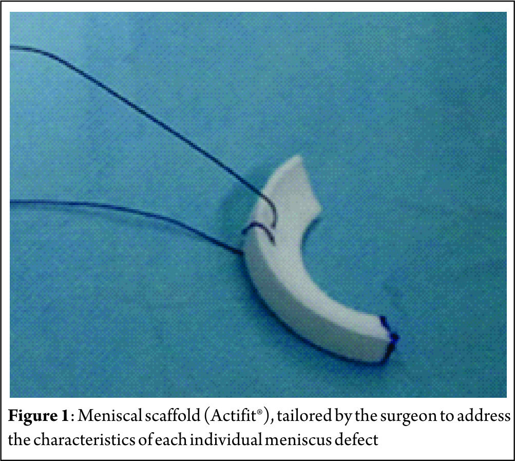
Collagen Meniscus Implant (CMI, Ivy Sports Medicine GmbH, Germany) – First published in 1997, CMI is a type-I collagen (isolated and purified from bovine Achilles tendon) scaffold (22) to which glycosaminoglycans are added. It has a meniscus-like shape, is implantable arthroscopically, and it is biocompatible and biodegradable. It has a microscopic porous structure that allows cellular ingrowth, induces differentiation and proliferation of fibrocartilaginous cells, leading to the creation of a meniscus-like tissue, concomitant with gradual resorption of the scaffold. Nevertheless, collagen scaffolds are fragile during the implant procedure, and have shown a decrease in size on follow-up magnetic resonance image (MRI) and arthroscopic second look follow-up. The second type of scaffold is Actifit® (Orteq, United Kingdom) that has been developed to overcome the perceived limitations of CMI related to difficulties in tissue handling with respect to suturing during implantation (Figure 1). Actifit® is composed of a slowly degrading polymer with polycaprolactone and urethane segments (23). Its structure seems to have better mechanical properties and is more resistant to sutures and loads as compared to CMI. The scaffold is 80% porous; the remaining 20% are made of a polymer with a low absorption rate. Degradation starts with hydrolysis of polycaprolactone segments, which lasts up to five years; the polyurethane segments are removed by macrophages and giants cells or integrated into surrounding tissues (24, 25).
Indications – Contraindications
When considering meniscal scaffolding, the surgeon should take into account several individual aspects, such as the patient’s age and weight, status of meniscal degeneration or concomitant conditions (such as axial malalignment and ligamentous insufficiency) (26). In this sense, several indications and contraindications have been developed as summarized in Table 1.

Preoperative Preparation
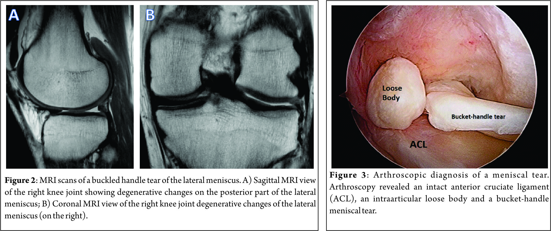
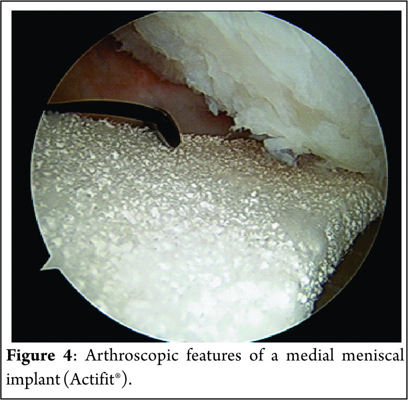
The preoperative imaging preparation usually involves radiography, MRI and, in some special cases, an arthro-computed tomography (arthro-CT). The radiographic imaging studies usually include bilateral comparison of weight-bearing radiographs (antero-posterior, lateral, Schuss or Rosenberg views). The MRI is usually performed to assess the cartilaginous structures status, quantify the meniscal damage, as well as the presence of bone marrow edema and/or meniscal extrusion (Figure 2). The arthro-CT scan may complement the MRI studies by assessing the meniscal volume and chondral damage (26). The imaging studies should be complemented with a comprehensive clinical examination of the knee. Special attention should be given to the knee ligament stability, as this has several implications in the meniscal surgery. In addition, diagnostic arthroscopy (Figure 3) may be performed to further assess the meniscal status and decide upon the best technique (26).
Surgical Technique
The procedure can be performed arthroscopically using the two standard anteromedial and anterolateral portals. The portals should be enlarged for an easier passage of the scaffold. The native remaining meniscus is thoroughly evaluated, and any torn or degenerative tissue is removed in order to leave a healthy and uniform meniscal rim, ensuring that the resulting defect site extends into the vascularized red-on-red or red-on-white zone of the meniscus. The meniscal rim is punctured in order to create vascular access channels. Gentle rasping of the synovial lining may further stimulate meniscal integration and tissue ingrowth. The exact size of the defect is measure with a flexible rod loaded in a rigid cannula starting at the posterior end of the lesion. The scaffold is measured and trimmed to the correct size on the sterile field of the operating environment (10% larger than in situ measurement to compensate for the shrinkage caused by suturing of the sponge-like material and to assure a snug optimal fit into the prepared defect). In order to achieve a perfect fit of the scaffold with the native meniscus at the anterior junction, the anterior side should be cut at an oblique angle of 30°-45°. The implant is inserted into the defect (Figure 4). Standard arthroscopic meniscal suturing techniques may be utilized for scaffold stabilization. The authors prefer “all-inside” vertical stitches placed every 4 to 5 mm to suture the scaffold along the periphery. The anterior and posterior scaffold extremities are fixed to the native remnant with horizontal stitches.
Concomitant Surgeries
Since other associated deficiencies (such as axial malalignment or ligamentous instability) may lead to poorer outcomes following meniscal surgery, these should be address in combination with the meniscal substitution (27). Anterior cruciate ligament insufficiency, if not addressed, may result in residual laxity, which may lead to an unfavorable meniscal healing environment. In this sense, ACL reconstruction has been performed along with the meniscal substitution in up to 67% of the patients (28-30). When performing concomitant ACL reconstruction, the meniscal bed should be firstly prepared and then the tibial and femoral tunnels may be drilled. After the tunnels are drilled, the ACL graft is passed through the tunnels and fixed at the femoral site, as the meniscal scaffold is inserted and sutured. Subsequently, the ACL graft is fixed at the tibial site with 20° of knee flexion (31). When uncorrected axial knee malalignments are found, these should be concomitantly or previously corrected. In a varus malalignment situation, a high tibial osteotomy may be performed to correct the malalignment. Special attention must be directed to the tibial slope and proper release of the medial collateral ligament should be performed. In valgus malalignments, if the deformity does not involve the tibial bone, osteotomy is done on the femoral side to avoid joint line obliquity (27).
Rehabilitation protocol
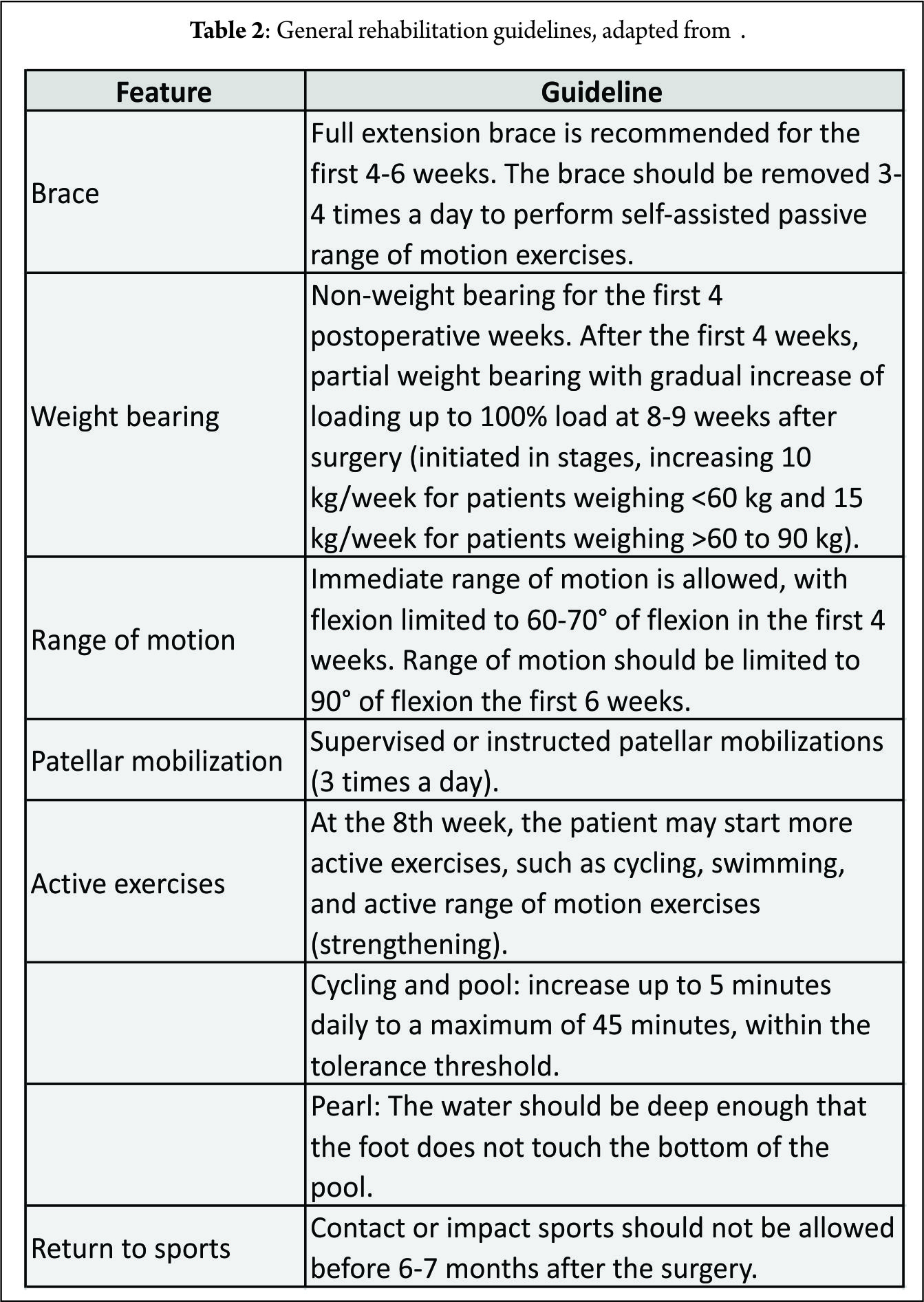
Patients are required to undergo a conservative rehabilitation program similar to that for a meniscal allograft. Special attention is required when the meniscal scaffold is implanted with concomitant ACL reconstruction or realignment osteotomy. In these cases, a rehabilitation program should be tailored to comply with the concomitant procedures postoperative particularities (26, 27). General guidelines for the rehabilitation program are presented in Table 3.
Clinical Studies
Although the literature contains clinical studies (33-35) that support the use of meniscal scaffold implantation for the treatment of irreparable meniscal tears, the quality of the studies is generally low, with lack of randomized trials and long-term follow-up to confirm clinical benefit and the most appropriate indications. Furthermore, long-term follow-up studies are required to verify the protective effect on the damaged joint compartment exerted by meniscal scaffold implantation.
A recent systematic literature review (35) analyzed results and indications for the treatment of meniscal loss. There has been an increase in publications regarding this topic recently, and the authors concluded that both CMI and Actifit seem to be safe and positive results have been shown for both scaffolds. Bulgheroni et al. (36) evaluated the safety and effectiveness of the polyurethane meniscal scaffold through clinical examination, MRI and arthroscopic second look, over a minimum two-year follow-up and showed no adverse reactions to the implant. The implant showed clear, hyperintense signal, sometimes irregular, and the chondral surface was preserved in all cases. At arthroscopic second look at 12 and 24 months, the scaffold was found to have an irregular morphology and to be slightly reduced in size. Zafagnini et al. (37), in a 10-year follow-up study, compared the medial collagen meniscus implant versus partial medial meniscectomy. The CMI group showed significantly lower visual analog scale scores for pain and higher objective International Knee Documentation Committee and Tegner index scores. Radiographic evaluation showed significantly less medial joint space narrowing in the CMI group compared to partial medial meniscectomy. No significant differences between groups were reported regarding Lysholm and Yulish scores. Another long-term study compared outcomes of CMI versus partial meniscectomy in patients with concomitant ACL reconstruction. The authors concluded that patients with chronic meniscal tears treated with medial CMI reported lower levels of post-operative pain compared to meniscectomy, while acute lesions treated with CMI showed less knee laxity at follow-up (38). The CMI when performed in the acute setting showed no additional benefits when compared to partial medial meniscectomy alone (28).
Zafagnini et al. (39), in a multi-center study, evaluated the clinical outcomes of 43 patients after lateral CMI implantation. They reported improvement of all clinical scores from baseline to follow-up evaluations. At the final follow-up, 58% of the patients reported activity levels comparable to their pre-injury values, with 95% patient reported satisfaction. A higher body mass index, the presence of concomitant procedures, and a chronic injury pattern were identified as potential negative prognostic factors.
As far as concomitant open-wedge high tibial osteotomies is concerned, Gelber et al. (40) found no short-term additional benefit when compared to partial meniscectomy and meniscal scaffolding.
Final Remarks and Future Directions
The menisci are known to be heterogeneous complex structures with segmental variations according to their anatomy, biology and function. The proper understanding on the different types of meniscal injuries (both traumatic and degenerative) and their pathophysiology and pathomechanics will assist the clinician in identifying the correct indications and contraindication for each type of lesion, preserving the meniscus whenever possible. The clinical application of meniscal scaffolds is limited to CMI and Actifit. In order to successfully implant these meniscus scaffolds, it is required an intact meniscal rim and sufficient meniscal tissue at the anterior and posterior meniscus horns to attach the scaffold. When in case of axial malalignments and/or ligament insufficiencies, these must be correct prior or during the scaffold implantation. The rehabilitation protocol should be tailored to address each patient’s individual characteristics, respect the chronobiology of the scaffold tissue integration and the progression within phases should be goal-based. Novel meniscal scaffolds have been developed for addressing total meniscus reconstruction with a functional meniscus replacement, mimicking the biology and mechanical properties of the native meniscus. These novel scaffolds may further protect the articular cartilage surface of the knee joint from the extensive damage after a total meniscectomy. A second generation of implants pre-cultured in vitro allows cell adhesion and extracellular matrix production and then are implanted into the meniscal defects which will probably follow as cell seeding as has been demonstrated to improve the mechanical properties and histological results. In the future, it may be possible to improve tissue formation in the meniscal scaffold using autologous cells (e.g., stem cells) and/or growth factors (e.g., platelet-rich plasma). This strategy may augment the tissue regeneration and improve clinical results. The use of mesenchymal stem cells may also enhance a greater promotion of intrinsic meniscal healing capacity. In addition, nanotechnology and gene therapy have emerged as potential options and have showed great potential for the treatment of meniscal lesions, however its translation into the clinical setting may take a few more years. Biofabrication of patient-specific meniscal scaffolds with a 3D printer from the advanced segmentation of menisci knee MRI datasets has been showing promising results in the laboratory setting. This novel technique will allow tailoring the meniscal scaffold to the patient-specific native characteristics of the knee.
References
1. Pereira H, Cengiz IF, Silva-Correia J, Cucciarini M, Gelber PE, Espregueira-Mendes J, Oliveira JM, Reis, RL. Histology-Ultrastructure-Biology. In: Hulet C, Pereira H, Peretti G, Denti, M, editors. Surgery of the Meniscus. Springer; 2016. p. 23-33.
2. Halewood C, Amis AA. Physiology: Biomechanics. In: Hulet C, Pereira H, Peretti G, Denti, M, editors. Surgery of the Meniscus. Springer; 2016. p. 35-45.
3. Heijink A, Gomoll AH, Madry H, Drobnič M, Filardo G, Espregueira-Mendes J, van Dijk CN. Biomechanical considerations in the pathogenesis of osteoarthritis of the knee. Knee Surg Sports Traumatol Arthrosc. 2012;20(3):423-35.
4. Ahmed A, Burke D. In-vitro of measurement of static pressure distribution in synovial joints—Part I: Tibial surface of the knee. J Biomech Eng. 1983;105(3):216-25.
5. Watanabe Y, Van Scyoc A, Tsuda E, Debski RE, Woo SL. Biomechanical function of the posterior horn of the medial meniscus: a human cadaveric study. J Orthop Sci. 2004;9(3):280-4.
6. Voloshin AS, Wosk J. Shock absorption of meniscectomized and painful knees: a comparative in vivo study. J Biomech Eng. 1983;5(2):157-61.
7. Abrams GD, Frank RM, Gupta AK, Harris JD, McCormick FM, Cole BJ. Trends in meniscus repair and meniscectomy in the United States, 2005-2011. Am J Sports Med. 2013:0363546513495641.
8. Verdonk PC, Dhollander AA, Tampere T, Verdonk R. Meniscus Substitution: Scaffolds, Allografts and Prosthetic Implants. In: Emans PJ, Peterson L, editors. Developing Insights in Cartilage Repair. Springer; 2014. p. 253-65.
9. Verdonk R. The meniscus: past, present and future. Knee Surg Sports Traumatol Arthrosc. 2011;19(2):145-6.
10. Di Matteo B, Tarabella V, Filardo G, Viganò A, Tomba P, Marcacci M. Thomas Annandale: the first meniscus repair. Knee Surg Sports Traumatol Arthrosc. 2013;21(9):1963-6.
11. Pereira H, Frias AM, Oliveira JM, Espregueira-Mendes J, Reis RL. Tissue engineering and regenerative medicine strategies in meniscus lesions. Arthroscopy. 2011;27(12):1706-19.
12. Cengiz IF, Pereira H, Pêgo JM, Sousa N, Espregueira‐Mendes J, Oliveira JM, Reis RL. Segmental and regional quantification of 3D cellular density of human meniscus from osteoarthritic knee. J Tissue Eng Regen Med. 2015. doi: 10.1002/term.2082.
13. Pereira H, Caridade S, Frias A, Silva-Correia J, Pereira D, Cengiz I, Mano JF, Oliveira JM, Espregueira-Mendes, J, Reis, RL. Biomechanical and cellular segmental characterization of human meniscus: building the basis for Tissue Engineering therapies. Osteoarthritis Cartilage. 2014;22(9):1271-81.
14. Cengiz IF, Pitikakis M, Cesario L, Parascandolo P, Vosilla L, Viano G, Oliveira, JM, Reis, RL. Building the Basis for Patient-Specific Meniscal Scaffolds: From Human Knee MRI to Fabrication of 3D Printed Scaffolds. Bioprinting. 2016.
15. Pereira H, Cengiz IF, Silva-Correia J, Oliveira JM, Reis RL, Espregueira-Mendes J. Human meniscus: from biology to tissue engineering strategies. In: Doral MN, Karlsson J, editos. Sports Injuries. Springer; 2015. p. 1089-102.
16. Yan L-P, Oliveira JM, Oliveira AL, Caridade SG, Mano JF, Reis RL. Macro/microporous silk fibroin scaffolds with potential for articular cartilage and meniscus tissue engineering applications. Acta Biomater. 2012;8(1):289-301.
17. Yan L-P, Silva-Correia J, Oliveira MB, Vilela C, Pereira H, Sousa RA, Mano JF, Oliveira AL, Oliveira JM, Reis, RL. Bilayered silk/silk-nanoCaP scaffolds for osteochondral tissue engineering: in vitro and in vivo assessment of biological performance. Acta Biomater. 2015;12:227-41.
18. Bacelar AH, Cengiz IF, Silva-Correia J, Sousa RA, Oliveira JM, L. RR. “Smart” Hydrogels in Tissue Engineering and Regenerative Medicine Applications. In: Khang G, editor. Handbook of Intelligent Scaffolds for Regenerative Medicine. Pan Stanford Publishing; 2015.
19. Buma P, Ramrattan N, van Tienen TG, Veth RP. Tissue engineering of the meniscus. Biomaterials. 2004;25(9):1523-32.
20. Reguzzoni M, Manelli A, Ronga M, Raspanti M, Grassi FA. Histology and ultrastructure of a tissue‐engineered collagen meniscus before and after implantation. J Biomed Mater Res B Appl Biomater. 2005;74(2):808-16.
21. Pereira H, Silva-Correia J, Oliveira J, Reis R, Espregueira-Mendes J. Future trends in the treatment of meniscus lesions: from repair to regeneration. In: Verdonk R, Espregueira-Mendes J, Monllau, JC, editors. Meniscal Transplantation. Springer; 2013. p. 103-12.
22. Stone KR, Steadman JR, Rodkey WG, Li S-T. Regeneration of Meniscal Cartilage with Use of a Collagen Scaffold. Analysis of Preliminary Data. J Bone Joint Surg Am. 1997;79(12):1770-7.
23. Verdonk R, Verdonk P, Huysse W, Forsyth R, Heinrichs E-L. Tissue ingrowth after implantation of a novel, biodegradable polyurethane scaffold for treatment of partial meniscal lesions. Am J Sports Med. 2011;39(4):774-82.
24. Van Minnen B, Van Leeuwen M, Kors G, Zuidema J, Van Kooten T, Bos R. In vivo resorption of a biodegradable polyurethane foam, based on 1, 4‐butanediisocyanate: A three‐year subcutaneous implantation study. J Biomed Mater Res A. 2008;85(4):972-82.
25. Zuidema J, van Minnen B, Span M, Hissink C, van Kooten T, Bos R. In vitro degradation of a biodegradable polyurethane foam, based on 1, 4‐butanediisocyanate: A three‐year study at physiological and elevated temperature. J Biomed Mater Res A. 2009;90(3):920-30.
26. Pujol N, Verdonk P. Actifit Polyurethane Meniscus Scaffold: Basic Science, Techniques, and Results. In: Hulet C, Pereira H, Peretti G, Denti, M, editors. Surgery of the Meniscus. Springer; 2016. p. 543-51.
27. Hinarejos P, Erggelet C, Monllau JC. Collagen Meniscus Implant: Basic Science, Technique and Results. In: Hulet C, Pereira H, Peretti G, Denti, M, editors. Surgery of the Meniscus. Springer; 2016. p. 531-42.
28. Rodkey WG, DeHaven KE, Montgomery WH, Baker CL, Beck CL, Hormel SE, Steadman R, Cole B, Briggs K. Comparison of the collagen meniscus implant with partial meniscectomy. J Bone Joint Surg Am. 2008;90(7):1413-26.
29. Monllau JC, Gelber PE, Abat F, Pelfort X, Abad R, Hinarejos P, et al. Outcome after partial medial meniscus substitution with the collagen meniscal implant at a minimum of 10 years’ follow-up. Arthroscopy. 2011;27(7):933-43.
30. Hirschmann M, Keller L, Hirschmann A, Schenk L, Berbig R, Lüthi U, et al. One-year clinical and MR imaging outcome after partial meniscal replacement in stabilized knees using a collagen meniscus implant. Knee Surg Sports Traumatol Arthrosc. 2013;21(3):740-7.
31. Monllau JC. Collagen Meniscal Implant (CMI). In: Verdonk R, Espregueira-Mendes J, Monllau, JC, editors. Meniscal Transplantation. Springer; 2013. p. 73-82.
32. Implant MCM. Rehabilitation program summary for patients, surgeons, and physiotherapists. Franklin Lakes, NJ, USA: ReGen Biologics, Inc.; 2006.
33. Grassi A, Zaffagnini S, Muccioli GMM, Benzi A, Marcacci M. Clinical outcomes and complications of a collagen meniscus implant: a systematic review. Int Orthop. 2014;38(9):1945-53.
34. Zaffagnini S, Grassi A, Muccioli GMM, Bonanzinga T, Nitri M, Raggi F, Ravazzolo J, Marcacci, M. MRI evaluation of a collagen meniscus implant: a systematic review. Knee Surg Sports Traumatol Arthrosc. 2015;23(11):3228-37.
35. Filardo G, Andriolo L, Kon E, de Caro F, Marcacci M. Meniscal scaffolds: results and indications. A systematic literature review. Int Orthop. 2015;39(1):35-46.
36. Bulgheroni P, Bulgheroni E, Regazzola G, Mazzola C. Polyurethane scaffold for the treatment of partial meniscal tears. Clinical results with a minimum two-year follow-up. Joints. 2013;1(4):161.
37. Zaffagnini S, Muccioli GMM, Lopomo N, Bruni D, Giordano G, Ravazzolo G, et al. Prospective Long-Term Outcomes of the Medial Collagen Meniscus Implant Versus Partial Medial Meniscectomy A Minimum 10-Year Follow-Up Study. Am J Sports Med. 2011;39(5):977-85.
38. Bulgheroni E, Grassi A, Bulgheroni P, Muccioli GMM, Zaffagnini S, Marcacci M. Long-term outcomes of medial CMI implant versus partial medial meniscectomy in patients with concomitant ACL reconstruction. Knee Surg Sports Traumatol Arthrosc. 2015;23(11):3221-7.
39. Zaffagnini S, Grassi A, Muccioli GMM, Holsten D, Bulgheroni P, Monllau JC, Berbig R, Lagae K, Crespo R, Marccaci M. Two-year clinical results of lateral collagen meniscus implant: a multicenter study. Arthroscopy. 2015;31(7):1269-78.
40. Gelber PE, Isart A, Erquicia JI, Pelfort X, Tey-Pons M, Monllau JC. Partial meniscus substitution with a polyurethane scaffold does not improve outcome after an open-wedge high tibial osteotomy. Knee Surg Sports Traumatol Arthrosc. 2015;23(1):334-9..
| How to Cite this article: Bastos R, Andrade R, Pereira H, Oliveira JM, Reis RL, Rodeo S, Espregueira-Mendes J. Meniscal Scaffolds in the Clinics: Present and future trends.Asian Journal of Arthroscopy Aug – Nov 2016;1(2):47-52 . |



Meniscectomy-Outcomes and Complications
Shantanu Sudhakar Patil, Sachin Ramchandra Tapasvi, Anshu Shekhar
Volume 1 | Issue 2 | Aug – Nov 2016 | Page 53-55.
Author: Shantanu Sudhakar Patil[1], Sachin Ramchandra Tapasvi[1], Anshu Shekhar[1].
[1] The Orthopaedic Speciality Clinic, 16 Status Chambers, 1221/A Wrangler Paranjpe Road, Pune 411004.
Address of Correspondence
Dr Sachin Ramchandra Tapasvi
The Orthopaedic Speciality Clinic, 16 Status Chambers, 1221/A Wrangler Paranjpe Road, Pune 411004
Email: stapasvi@gmail.com
Abstract
The menisci, once considered expendable remnants have been conclusively proven to be of extreme vitality in the biomechanics and biology of the knee joint. Though meniscus repair is being increasingly performed to preserve knee function, not all tears are amenable to repair and partial meniscectomy in such cases is an acceptable treatment option. The poor outcomes following partial meniscectomy are due to the shrinking of contact areas and rise in peak stresses. These changes and their consequences are more pronounced in the lateral compartment of the knee. Pre-existing chondral damage, instability and higher BMI compound the problem.
Key words: Meniscus, meniscectomy, meniscus repair, arthritis.
Introduction
The menisci of the knee joint are fibrocartilagenous semilunar tissues that perform a critical function of stabilising the joint and aiding in efficient load transfer as a shock absorber. Though once considered vestigial and hence disposable, the role of healthy menisci in delaying the normal attrition of the articular cartilage cannot be understated. Meniscectomy was thought to be a benign procedure and as late as 1975 [1] the importance of doing a complete removal was being reiterated. The functions of the meniscus were recognised much earlier [2] and eventually the potential harms of its excision were gaining attention. Meniscal tears are one of the commonest injuries of the knee, for which treatment is sought, with an incidence rate of 61 per 100000 population per year.[3] Most acute tears are commoner in younger patients, with the medial meniscus affected at a 2:1 ratio with the lateral side. The acute tears are described as per their orientation and extent along the meniscus. They are usually classified as vertical longitudinal, oblique, circumferential, complex, transverse or radial, and horizontal cleavage tears. Radial tears of the posteromedial compartment are the most frequently seen tears and vertical longitudinal tears are most often associated with acute ACL injury. Degenerative tears have a varied pattern and are complex in their morphology.[4]. The direction of the meniscus tears is explained by the orientation of collagen fibrils within the structure. The cross section of the meniscus reveals three distinct layers: a superficial thin layer on both tibial and femoral surfaces; a lamellar layer below this with the fibrils arranged in a radial manner and a main central region where the fibrils are orientated in a circular manner. The circular arrangement of the collagen bundles explains why majority of the tears have a longitudinal orientation. [5](Fig. 1). With our growing understanding of the anatomy , vasculature, biomechanics and the biology of the meniscus, and with improved arthroscopic techniques and instrumentation, the goal of management of meniscal tears has shifted towards achieving repair. However, not all tears are amenable to repair and at least a partial meniscectomy might be indicated to alleviate the patients symptoms. We will take a look at the outcomes and complications of arthroscopic meniscectomy in this article.
Sequelae of articular cartilage changes following Meniscectomy
The effects of meniscectomy on the stability and pressures inside the knee joint were studied using pressure sensitive films in cadavers. Medial meniscectomy caused the contact areas to shrink by almost 75% leading to more than twofold increase in peak contact pressures. [6] The articular cartilage responds unfavourably to the higher loads, with disruption of the proteoglycan matrix, causing swelling and inflammation throughout the joint. The heightened catabolic state with increased hydration leads to breakdown of the collagen matrix, thus accelerating the normal wear and tear within the joint.[7]
Radiological changes:
The radiological changes in the knee joint following medial meniscectomy are well documented.[8]Joint space narrowing, flattening of the marginal part of the medial femoral condyle and sclerosis of the articulating condyles is seen. These radiological signs were indicative of early osteoarthritic changes in the knee. Multiple clinical and radiological studies have documented these sequelae, but the correlation between the symptoms of the patient and severity of these changes is not always seen in the results. It is not easy to determine the correlations as many reports have studied the consequences after an open meniscectomy. Moreover, a meniscal tear rarely presents in isolation and the concomitant ligament or articular injuries play a role in subsequent degeneration and development of Osteoarthritis.
Partial Versus Total meniscectomy
With the advent of arthroscopic surgery and advances in instrumentation for the various surgical procedures, it was possible to resect only the offending parts of the torn meniscus. It is uncommon these days to perform a total resection, with partial meniscectomy being the more widely reported procedure. Once a meniscal tear is identified and deemed unsuitable for repair, a meniscectomy is the recommended surgical option. The basic principles for this were described by Metcalf. They are as follows: Remove all mobile fragments; Avoid sudden changes in rim contour; a perfectly smooth rim is unnecessary as some remodelling may occur; re-evaluate the tear often with a probe; Avoid damage to the meniscus-capsular junction to avoid the loss of hoop stresses; Use both manual and motorized instruments to maximize efficiency and when uncertain if an area should be resected, err on the side of leaving more meniscus intact rather than compromising biomechanical properties[9]. Salata in a meta-analysis showed the significantly higher risk of developing radiographic OA in the patients undergoing total meniscectomy as compared to the partial meniscectomy. [10] Though the patients with either partial or total meniscectomy report similar early clinical results, there was no significant difference in the radiographic outcomes at the final 7.8 years average follow-up[11]. Only 68% of the patients who had undergone a total meniscectomy and followed up for up to 30 years showed good or excellent results while at least 2/3rd had some post-operative symptoms.[12] There exists a direct correlation with the meniscal tissue left behind and peak contact stress on the tibial surfaces following partial resections[13]. A finite element study quantifying the amount of resected meniscus to peak pressures showed that with as little as 20% resection of meniscus, a detrimental increase of forces is seen which may hasten the osteoarthritic changes. Maximum shear stress in the articular cartilage is seen with 65% partial meniscectomy[14]. The orientation of collagen fibril bundles within the meniscus determines the development of hoop strains as they are axially loaded. A radial tear disrupts the continuity of the circularly oriented fibrils and thus prevents the hoop strains from forming, causing dysfunction of the meniscus. A horizontal or vertical tear will not disrupt this continuity, preserving the load-bearing and shock-bearing function of the meniscus.[15] This needs to be borne in mind while determining the extent of the meniscectomy. The medial and lateral tibio-femoral articulations are anatomically different and the absence of menisci which afford a degree of congruity can lead to increased point loading and higher contact pressures. This is more prominent on the lateral side where a convex lateral femoral condyle articulates on a flat or convex tibial plateau. This translates to poorer outcomes with lateral meniscectomy as compared to medial as reported in multiple studies. Patients with a lateral meniscectomy have a much higher functional deterioration and increased instability than the medial meniscectomy patients[11,3,16].
Influence of other concomitant factors
The ACL-deficient knee with a meniscal tear has a significantly higher radiographic grade changes after meniscectomy as compared with ACL-intact knees.[17] Consequences of meniscectomy in an unstable knee are worsened by the combination of higher contact forces inducing early pathological changes due to the elevated shear stresses within the articular cartilage.[18] The presence of pre-existing chondral damage at the time of meniscectomy predisposes the knee to a significant increase in development of OA leading to poor clinical outcomes. However, contradicting findings have also been reported with there being no significant changes in knee functions and activity level following the meniscectomy. [19]. Chondral lesions can cause similar symptoms as that of a meniscal tear and meniscectomy may not fully alleviate the patients’ complaints, thus leading to poor outcomes. Degenerative tears are more often seen in older subjects with varus alignment. However, the evidence that meniscectomy in this group leads to higher rate of radiographic OA is not conclusive. These patients do show a decreased level of activity along with poorer outcomes based on subjective and functional measures following the surgery.[19, 20]While there is consensus about patients with increased BMI predisposing to a higher risk of OA post meniscectomy, the exact level of BMI that placed the patient at risk is not conclusive.[11], [21].
Complications
These can be classified as those related to knee arthroscopy in general and those associated specifically with arthroscopic partial meniscectomy. In the hands of an experienced arthroscopy surgeon, the complication rates were low. (1.78% and 1.48% for medial and lateral meniscectomy) [22], [23]. Some of the enumerated complications include instrument failure or breakage, injuries to nerves and blood vessels, accidental damage to chondral surfaces and ligament injury. Instrument failure rates have dropped from 18.1% to 2.9% over the years, due to improvement in surgical techniques, better designs as well as better skill levels of the surgeons[22]. The medial collateral ligament may get injured due to excessive valgus forces while attempting access to the medial compartment. Similarly, nerves and vessels may get damaged during insertion of sharp instruments. Improper and clumsy handling of instruments during the surgery can gouge the articular surface causing damage. Incomplete removal of the torn pieces can cause persistent pain along with coexistent knee pathology. Proper adherence to basic principles of partial meniscectomy can help avoid all these complications.
Conclusion
There exist a large number of studies which have studied the consequences of meniscectomy as a surgical procedure. Many of these have incomplete or inaccurate information along with varying heterogeneous criteria for evaluation of outcomes. The functional and clinical outcomes do not necessarily match the radiological outcomes in most of the studies. The multiple imaging modalities add to data which is not uniform for evaluation. This lack of homogenous data and lack of standardization of methodological issues, makes it difficult to conclude if the findings represent true differences or are simply artefact related to measurement bias or other errors. It is probably safe to conclude that a minimally invasive procedure with attention to sparing bulk of meniscal tissue seems to reduce the subsequent incidence of arthritic changes, as compared with open invasive and radical procedures..
References
1. Hughston, J.C., A simple meniscectomy. J Sports Med, 1975. 3(4): p. 179-87.
2. King, D., The healing of semilunar cartilages. 1936. Clin Orthop Relat Res, 1990(252): p. 4-7.
3. Jones, J.C., et al., Incidence and risk factors associated with meniscal injuries among active-duty US military service members. J Athl Train, 2012. 47(1): p. 67-73.
4. Pauli, C., et al., Macroscopic and histopathologic analysis of human knee menisci in aging and osteoarthritis. Osteoarthritis Cartilage, 2011. 19(9): p. 1132-41.
5. Petersen, W. and B. Tillmann, Collagenous fibril texture of the human knee joint menisci. Anat Embryol (Berl), 1998. 197(4): p. 317-24.
6. Baratz, M.E., F.H. Fu, and R. Mengato, Meniscal tears: the effect of meniscectomy and of repair on intraarticular contact areas and stress in the human knee. A preliminary report. Am J Sports Med, 1986. 14(4): p. 270-5.
7. Lanzer, W.L. and G. Komenda, Changes in articular cartilage after meniscectomy. Clin Orthop Relat Res, 1990(252): p. 41-8.
8. Fairbank, T.J., Knee joint changes after meniscectomy. J Bone Joint Surg Br, 1948. 30b(4): p. 664-70.
9. Metcalf, R.W., Arthroscopic meniscal surgery., in Operative Arthroscopy., M. JB, Editor. 1991, Raven Press: New York. p. pp. 203–236.
10. Salata, M.J., A.E. Gibbs, and J.K. Sekiya, A systematic review of clinical outcomes in patients undergoing meniscectomy. Am J Sports Med, 2010. 38(9): p. 1907-16.
11. Hede, A., E. Larsen, and H. Sandberg, Partial versus total meniscectomy. A prospective, randomised study with long-term follow-up. J Bone Joint Surg Br, 1992. 74(1): p. 118-21.
12. Tapper, E.M. and N.W. Hoover, Late results after meniscectomy. J Bone Joint Surg Am, 1969. 51(3): p. 517-26 passim.
13. Ihn, J.C., S.J. Kim, and I.H. Park, In vitro study of contact area and pressure distribution in the human knee after partial and total meniscectomy. Int Orthop, 1993. 17(4): p. 214-8.
14. Vadher, S.P., et al., Finite element modeling following partial meniscectomy: effect of various size of resection. Conf Proc IEEE Eng Med Biol Soc, 2006. 1: p. 2098-101.
15. Jones, R.S., et al., Direct measurement of hoop strains in the intact and torn human medial meniscus. Clin Biomech (Bristol, Avon), 1996. 11(5): p. 295-300.
16. Petty, C.A. and J.H. Lubowitz, Does arthroscopic partial meniscectomy always cause arthritis? Sports Med Arthrosc, 2012. 20(2): p. 58-61.
17. Burks, R.T., M.H. Metcalf, and R.W. Metcalf, Fifteen-year follow-up of arthroscopic partial meniscectomy. Arthroscopy, 1997. 13(6): p. 673-9.
18. McDermott, I.D. and A.A. Amis, The consequences of meniscectomy. J Bone Joint Surg Br, 2006. 88(12): p. 1549-56.
19. Rockborn, P. and J. Gillquist, Long-term results after arthroscopic meniscectomy. The role of preexisting cartilage fibrillation in a 13 year follow-up of 60 patients. Int J Sports Med, 1996. 17(8): p. 608-13.
20. Chatain, F., et al., The natural history of the knee following arthroscopic medial meniscectomy. Knee Surgery, Sports Traumatology, Arthroscopy, 2000. 9(1): p. 15-18.
21. Englund, M. and L.S. Lohmander, Risk factors for symptomatic knee osteoarthritis fifteen to twenty-two years after meniscectomy. Arthritis Rheum, 2004. 50(9): p. 2811-9.
22. Small, N.C., Complications in arthroscopic surgery performed by experienced arthroscopists. Arthroscopy, 1988. 4(3): p. 215-21.
23. Allum, R., Complications of arthroscopy of the knee. J Bone Joint Surg Br, 2002. 84(7): p. 937-45.
24. Papalia, R., et al., Meniscectomy as a risk factor for knee osteoarthritis: a systematic review. Br Med Bull, 2011. 99: p. 89-106.
(Abstract) (Full Text HTML) (Download PDF)
Menisci: Structure and Function
Ankit Chawla, Amite Pankaj Aggarwal
Volume 1 | Issue 2 | Aug – Nov 2016 | Page 3-7
Author: Ankit Chawla[1], Amite Pankaj Aggarwal[1]
[1] Unit of Joint Replacement, Arthroscopy and Orthopaedics, Fortis Hospital, Shalimar Bagh, New Delhi, India.
Address of Correspondence
Dr. Amite Pankaj Aggarwal
Fortis Hospital Shalimar Bagh
New Delhi, India.
Email: amitepankaj@gmail.com.
Abstract
Meniscal injuries are recognized as a cause of significant musculoskeletal morbidity. The menisci are vital for the normal function and long-term health of the knee joint. And loss of a meniscus increases the risk of subsequent development of degenerative changes in the knee. A review of anatomy and ultrastructure of the meniscus, and its relationship to normal function in terms of load transmission, shock absorption, joint stability, lubrication and nutrition is a necessary prerequisite to understanding pathologies associated with the knee.
Keywords: Meniscus, Medial meniscus, lateral meniscus, Anatomy, Function.
Introduction
The word meniscus comes from the Greek word me-niskos, meaning “crescent,” diminutive of me-ne-, meaning “moon.” The menisci are semilunar discs of fibrocartilaginous tissue which are vital for the normal biomechanics and long-term health of the knee joint [1]. The characteristic shape of the lateral and medial menisci is attained between the 8th and 10th week of gestation. They arise from a condensation of the intermediate layer of mesenchymal tissue to form attachments to the surrounding joint capsule[2,3].
Gross Anatomy
These crescent-shaped wedges of fibrocartilage are located on the medial and lateral aspects of the knee joint (Fig. 1A,1B). The peripheral, vascular border of each meniscus is thick, convex, and attached to the joint capsule. The innermost border tapers to a thin free edge. The superior surfaces of menisci are concave, enabling effective articulation with their respective convex femoral condyles. The inferior surfaces are flat to accommodate the tibial plateau [4,5].
Medial Meniscus
The medial meniscus is a C-shaped structure larger in radius than the lateral meniscus, with the posterior horn being wider than the anterior. The anterior horn is attached firmly to the tibia anterior to the intercondylar eminence and to the anterior cruciate ligament. The posterior horn is anchored immediately in front of the attachments of the posterior cruciate ligament posterior to the intercondylar eminence. Its entire peripheral border is firmly attached to the medial capsule and through the coronary ligament to the upper border of the tibia. At its midpoint, the medial meniscus is more firmly attached to the femur through a condensation in the joint capsule known as the deep medial collateral ligament [5]. The transverse, or “intermeniscal,” ligament is a fibrous band of tissue that connects the anterior horn of the medial meniscus to the anterior horn of the lateral meniscus [5,6].
Lateral Meniscus
The lateral meniscus is more circular in form, covering up to two thirds of the articular surface of the underlying tibial plateau [7]. The anterior horn is attached to the tibia medially in front of the intercondylar eminence, whereas the posterior horn inserts into the posterior aspect of the intercondylar eminence and in front of the posterior attachment of the medial meniscus. The lateral meniscus is loosely attached to the capsular ligament; however, these fibers do not attach to the lateral collateral ligament. The posterior horn of the lateral meniscus attaches to the inner aspect of the medial femoral condyle via the anterior and posterior meniscofemoral ligaments of Humphrey and Wrisberg, respectively, which originate near the origin of the PCL (Fig. 1A) [8]. Their estimated prevalence is 74 % for Humphrey ligament, 69 % for Wrisberg ligament, and both ligaments found together in around 50 % of knees [9]. The lateral meniscus is smaller in diameter, thicker in periphery, wider in body, and more mobile than the medial meniscus.
Extracellular matrix and cellularity
Considering composition by wet weight, the meniscus has high water content (72 %). The remaining 28 % consists of an organic component, mostly ECM and cells.10 Collagens comprise the majority (75 %) of the organic matter, followed by GAGs (17 %), DNA (2 %), adhesion glycoproteins (<1 %), and elastin (<1 %) [10,11]. These proportions vary according to age, injury, or pathological conditions [12]. Collagen is the main fibrillar component of the meniscus. Different collagen types exist in various quantities in each region of meniscus. In the red–red zone, type I collagen is predominant (80 % composition in dry weight). In the white–white zone, 60 % is type II collagen and 40 % is type I collagen [13]. The major orientation of collagen fibers in the meniscus is circumferential; radial fibers and perforating fibers also are present.(Fig. 3) [13]. Proteoglycans are heavily glycosylated molecules that constitute a major component of the meniscus ECM [14]. These molecules are comprised of a core protein which is decorated with glycosaminoglycans (GAGs). The main types of GAGs found in normal human meniscal tissue are chondroitin 6 sulfate (60%), dermatan sulfate(20-30%), chondroitin 4 sulfate (10-20%), and keratin sulfate(15%) [15]. Their main function is to enable the meniscus to absorb water, whose confinement supports the tissue under compression [10]. Adhesion glycoproteins are also important components of the meniscus matrix, as they serve as a link between ECM components and cells [16]. The main adhesion glycoproteins present in the human meniscus are fibronectin, thrombospondin, and collagen VI [16,17]. Outer zone cells have an oval, fusiform shape and are similar in appearance and behaviour to fibroblasts, described as fibroblast-like cells [18]. The matrix surrounding the cells is mainly comprised of type I collagen, with small percentages of glycoproteins and collagen types III and V present. In contrast, cells in the inner portion have rounded appearance and are embedded in an ECM comprising largely type II collagen intermingled with a smaller but significant amount of type I collagen and higher concentration of GAGs [18]. This relative abundance of collagen type II and aggrecan in the inner region is more reminiscent of hyaline articular cartilage. Therefore, cells in this region are classified as fibrochondrocytes or chondrocyte like cells. In summary, cell phenotype and ECM composition render the outer portion of the meniscus akin to fibrocartilage, while the inner portion possesses similar, but not identical, traits to articular cartilage [19,20].
Vascularity and Innervation
The vascular supply to the medial and lateral menisci originates predominantly from the lateral and medial geniculate vessels (both inferior and superior). Branches from these vessels give rise to a perimeniscal capillary plexus within the synovial and capsular tissue. (Fig. 2) Radial branches from the plexus enter the meniscus at intervals, with a richer supply to the anterior and posterior horns. Vessels supplying the body are limited to the meniscus periphery with a variable penetration of 10–30 % for medial meniscus and 10–25 % for lateral one. This has important implication for meniscal healing [21]. The remaining portion of each meniscus (65% to 75%) receives nourishment from synovial fluid via diffusion or mechanical pumping (ie, joint motion) [22, 23]. The knee joint is innervated by the posterior articular branch of the posterior tibial nerve and the terminal branches of the obturator and femoral nerves. The lateral portion of the capsule is innervated by the recurrent peroneal branch of the common peroneal nerve. These nerve fibers penetrate the capsule and follow the vascular supply to the peripheral portion of the menisci and the anterior and posterior horns, where most of the nerve fibers are concentrated. The inner menisci core has no nerve fibers [21].
Biomechanical Function
The biomechanical function of the meniscus is a reflection of the gross and ultrastructural anatomy and of its relationship to the surrounding intra-articular and extra-articular structures. The meniscus withstands many different forces such as shear, tension, and compression. It also plays a crucial role in load-bearing, load transmission, shock absorption, stability, propioception as well as lubrication and nutrition of articular cartilage [24-27]. They also serve to decrease contact stresses and increase contact area and congruity of the knee [28,29].
Meniscal Biomechanics
The biomechanical properties of the knee meniscus are appropriately tuned to withstand the forces exerted on the tissue. Many studies have helped to quantify the properties of the tissue both in humans and in animal models. According to these studies, the meniscus resists axial compression with an aggregate modulus of 100-150 kPa [30]. The tensile modulus of the tissue varies between the circumferential and radial directions; it is approximately 100-300 MPa circumferentially and 10 fold lower than this radially [31]. Finally, the shear modulus of the meniscus is approximately 120 kPa [31]. The contact forces on the meniscus within the human knee joint have been mapped. It has been calculated that the intact menisci occupy approximately 60% of the contact area between the articular cartilage of the femoral condyles and the tibial plateau, while they transmit >50% of the total axial load applied in the joint [32,33]. However, these percentages are highly dependent on degree of knee flexion and tissue health. For every 30o of knee flexion, the contact surface between the two knee bones decreases by 4% [34]. When the knee is in 90o of flexion the applied axial load in the joint is 85% greater than when it is in 0o of flexion [33]. In full knee flexion, the lateral meniscus transmits 100% of the load in the lateral knee compartment, whereas the medial meniscus takes on approximately 50% of the medial load [29]. Studies confirm that there is a significant difference in segmental motion during flexion between the medial and lateral menisci. The anterior and posterior horn lateral meniscus ratio is smaller and indicates that the meniscus moves more as a single unit [35]. Alternatively, the medial meniscus (as a whole) moves less than the lateral meniscus, displaying a greater anterior to posterior horn differential excursion. Thompson et al found that the area of least meniscal motion is the posterior medial corner, where the meniscus is constrained by its attachment to the tibial plateau by the meniscotibial portion of the posterior oblique ligament, which has been reported to be more prone to injury [35,36]. A reduction in the motion of the posterior horn of the medial meniscus is a potential mechanism for meniscal tears, with a resultant “trapping” of the fibrocartilage between the femoral condyle and the tibial plateau during full flexion. The greater differential between anterior and posterior horn excursion may place the medial meniscus at a greater risk of injury [35]. The differential of anterior horn to posterior horn motion allows the menisci to assume a decreasing radius with flexion, which correlates to the decreased radius of curvature of the posterior femoral condyles [35]. This change of radius allows the meniscus to maintain contact with the articulating surface of both the femur and the tibia throughout flexion.
Load Transmission
Fairbank described the increased incidence and predictable degenerative changes of the articular surfaces in completely meniscectomized knees [37]. Weightbearing produces axial forces across the knee, which compress the menisci, resulting in “hoop” (circumferential) stresses [38]. Hoop stresses are generated as axial forces and converted to tensile stresses along the circumferential collagen fibers of the meniscus. Firm attachments by the anterior and posterior insertional ligaments prevent the meniscus from extruding peripherally during load bearing [39]. Medial meniscectomy decreases contact area by 50% to 70% and increases contact stress by 100%. Lateral meniscectomy decreases contact area by 40% to 50% but dramatically increases contact stress by 200% to 300% because of the relative convex surface of the lateral tibial plateau [40,41]. This significantly increases the load per unit area and may contribute to accelerated articular cartilage damage and degeneration [42].
Shock absorption
The menisci play a vital role in attenuating the intermittent shock waves generated by impulse loading of the knee with normal gait [43,44]. Voloshin and Wosk showed that the normal knee has a shock-absorbing capacity about 20% higher than knees that have undergone meniscectomy [38]. As the inability of a joint system to absorb shock has been implicated in the development of osteoarthritis, the meniscus would appear to play an important role in maintaining the health of the knee joint [45]
Joint stability
The geometric structure of the menisci provides an important role in maintaining joint congruity and stability. The superior surface of each meniscus is concave, enabling effective articulation between the convex femoral condyles and flat tibial plateau. When the meniscus is intact, axial loading of the knee has a multidirectional stabilizing function, limiting excess motion in all directions [46]. The studies for effects of meniscectomy on joint laxity for anteroposterior and varus-valgus motions and rotation have indicated indicated that the effect on joint laxity depends on whether the ligaments of the knee are intact and whether the joint is bearing weight. In the presence of intact ligamentous structures, excision of the menisci produces small increases in joint laxity. In an anterior cruciate ligament–deficient knee, medial meniscectomy has been shown to increase tibial translation by 58% at 90o, whereas primary anterior and posterior translations were not affected by lateral meniscectomy [47]. Shoemaker and Markolf demonstrated that the posterior horn of the medial meniscus is the most important structure resisting an anterior tibial force in the ACL-deficient knee. [48] Recently, Musahl et al reported that the lateral meniscus plays a role in anterior tibial translation during the pivot-shift maneuver [49].
Joint Nutrition and Lubrication
The menisci may also play a role in the nutrition and lubrication of the knee joint. The mechanics of this lubrication remains unknown; the menisci may compress synovial fluid into the articular cartilage, which reduces frictional forces during weightbearing [50]. There is a system of microcanals within the meniscus located close to the blood vessels, which communicates with the synovial cavity; these may provide fluid transport for nutrition and joint lubrication [51,52].
Conclusions
Mechanoreceptors have been identified in the anterior and posterior horns of the menisci, middle and outer third of the meniscus. The identification of these neural elements indicates that the menisci are capable of detecting proprioceptive information (joint motion and position) in the knee joint, thus playing an important afferent role in the sensory feedback mechanism of the knee [53,54].
References
1. McDermott ID, Masouros SD, Amis AA. Biomechanics of the menisci of the knee. Curr Orthopaed.2008 ;22:193–201
2. Gardner E, O’Rahilly R. The early development of the knee joint in staged human embryos. J Anat.1968;102:289-299.
3. Gray DJ, Gardner E. Pre-natal development of the human knee and superior tibial fibula joints. Am J Anat.1950;86:235-288.
4. Bullough PG, Vosburgh F, Arnoczky SP, et al. The menisci of the knee. In: Insall JN, ed. Surgery of the Knee. New York, NY: Churchill Livingstone; 1984:135-149.
5. Warren RF, Arnoczky SP, Wickiewiez TL. Anatomy of the knee. In: Nicholas JA, Hershman EB, eds. The Lower Extremity and Spine in Sports Medicine. St. Louis: Mosby; 1986:657-694.
6. Johnson DL, Swenson TD, Harner CD. Arthroscopic meniscal transplantation: anatomic and technical considerations. Presented at: Nineteenth Annual Meeting of the American Orthopaedic Society for Sports Medicine; July 12-14, 1993; Sun Valley, ID.
7. Arnoczky SP, Adams ME, DeHaven KE, Eyre DR, Mow VC. The meniscus. In: Woo SL-Y, Buckwalter J, eds. Injury and Repair of Musculoskeletal Soft Tissues. Park Ridge, IL: American Academy of Orthopaedic Surgeons; 1987:487-537.
8. Johnson DL, Swenson TM, Livesay GA, Aizawa H, Fu FH, Harner CD. Insertion-site anatomy of the human menisci: gross, arthroscopic, and topographical anatomy as a basis for meniscal transplantation. Arthroscopy.1995;11:386-394.
9. Heller L, Langman J. The meniscofemoral ligaments of the human knee. J Bone Joing Surg Br.1964;46 :307-313.
10. Makris EA, Hadidi P, Athanasiou KA. The knee meniscus: structure-function, pathophysiology, current repair techniques, and prospects for regeneration. Biomaterials. 2011 Oct;32(30):7411-31.
11. Herwig J, Egner E, Buddecke E. Chemical changes of human knee joint menisci in various stages of degeneration. Ann Rheum Dis. 1984 Aug;43(4):635-40.
12. Sweigart MA, Athanasiou KA. Toward tissue engineering of the knee meniscus. Tissue Eng. 2001 Apr;7(2):111-29.
13. Cheung HS. Distribution of type I, II, III and V in the pepsin solubilized collagens in bovine menisci. Connect Tissue Res. 1987;16(4):343-56.
14. Beaupre A, Choukroun R, Guidouin R, Carneau R, Gerardin H. Knee menisci: correlation between microstructure and biomechanics. Clin Orthop Relat Res.1986 ;208 :72-75.
15. Assimakopoulos AP, Katonis PG, Agapitos MV, Exarchou EI. The innervations of the human meniscus. Clin Orthop Relat Res.1992;275:232-236.
16. Miller RR, McDevitt CA. Thrombospondin in ligament, meniscus and intervertebral disc. Biochim Biophys Acta. 1991 Nov 14;1115(1):85-8
17. McDevitt CA, Webber RJ. The ultrastructure and biochemistry of meniscal cartilage. Clin Orthop Relat Res. 1992; 252:8–18.
18. Verdonk PC, Forsyth RG, Wang J et al (2005) Characterisation of human knee meniscus cell phenotype. Osteoarthritis Cartilage 13:548–560.
19. Melrose J, Smith S, Cake M, Read R, Whitelock J. Comparative spatial and temporal localisation of perlecan, aggrecan and type I, II and IV collagen in the ovine meniscus: an ageing study. Histochem Cell Biol. 2005; 124:225–35.
20. Hellio Le Graverand MP, Ou Y, Schield-Yee T, Barclay L, Hart D, Natsume T, et al. The cells of the rabbit meniscus: their arrangement, interrelationship, morphological variations and cytoarchitecture. J Anat. 2001; 198:525–35.
21. Brian D, Mackenzie WG, Shim SS, Leung G (1985) The vascular and nerve supply of the human meniscus. Arthroscopy 1:58–62.
22. Meyers E, Zhu W, Mow V. Viscoelastic properties of articular cartilage and meniscus. In: Nimni M, ed. Collagen: Chemistry, Biology and Biotechnology. Boca Raton, FL: CRC; 1988.
23. Mow V, Fithian D, Kelly M. Fundamentals of articular cartilage and meniscus biomechanics. In: Ewing JW, ed. Articular Cartilage and Knee Joint Function: Basic Science and Arthroscopy New York, NY: Raven Press; 1989:1-18.
24. Cameron HU, Macnab I. The structure of the meniscus of the human knee joint. Clin Orthop Relat Res. 1972; 89:215–9.
25. Newman AP, Anderson DR, Daniels AU, Dales MC. Mechanics of the healed meniscus in a canine model. Am J Sports Med. 1989; 17:164–75.
26. Zhu W, Chern KY, Mow VC. Anisotropic viscoelastic shear properties of bovine meniscus. Clin Orthop Relat Res. 1994; 306:34–45.
27. Tissakht M, Ahmed AM, Chan KC. Calculated stress-shielding in the distal femur after total knee replacement corresponds to the reported location of bone loss. J Orthop Res. 1996; 14:778–85.
28. Kettelkamp DB, Jacobs AW. Tibiofemoral contact area: determination and implications. J Bone Joint Surg Am. 1972;54:349-356.
29. Walker PS, Erkman MJ. The role of the meniscus in force transmission across the knee. Clin Orthop Relat Res. 1975;109:184-192.
30. Sweigart MA, Zhu CF, Burt DM, DeHoll PD, Agrawal CM, Clanton TO, et al. Intraspecies and interspecies comparison of the compressive properties of the medial meniscus. Ann Biomed Eng. 2004; 32:1569–79.
31. Fithian DC, Kelly MA, Mow VC. Material properties and structure-function relationships in the menisci. Clin Orthop Relat Res. 1990; 252:19–31.
32. Fukubayashi T, Kurosawa H. The contact area and pressure distribution pattern of the knee. A study of normal and osteoarthrotic knee joints. Acta Orthop Scand. 1980; 51:871–9.
33. Ahmed AM, Burke DL. In-vitro measurement of static pressure distribution in synovial joints—Part I: Tibial surface of the knee. J Biomech Eng. 1983; 105:216–25.
34. Walker PS, Hajek JV. The load-bearing area in the knee joint. J Biomech. 1972; 5:581–9.
35. Thompson WO, Thaete FL, Fu FH, Dye SF. Tibial meniscal dynamics using three-dimensional reconstruction of magnetic resonance imaging. Am J Sports Med.1991;19:210-216.
36. Ricklin P, Ruttimann A, Del Bouno MS. Diagnosis, Differential Diagnosis and Therapy. 2nd ed. Stuttgart, Germany: Verlag Georg Thieme; 1983
37. Fairbank TJ. Knee joint changes after meniscectomy. J Bone Joint Surg Br.1948;30:664-670.
38. Voloshin AS, Wosk J. Shock absorption of meniscectomized and painful knees: a comparative in vivo study. J Biomed Eng.1983 ;5 :157-161.
39. Krause WR, Pope MH, Johnson RJ, Wilder DG. Mechanical changes in the knee after meniscectomy. J Bone Joint Surg Am. 1976;58:599-604.
40. Fukubayashi T, Kurosawa H. The contact area and pressure distribution pattern of the knee: a study of normal and osteoarthritic knee joints. Acta Orthop Scand.1980;51:871-879.
41. Kettelkamp DB, Jacobs AW. Tibiofemoral contact area: determination and implications. J Bone Joint Surg Am.1972;54:349-356.
42. Jones RE, Smith EC, Reisch JS. Effects of medial meniscectomy in patients older than forty years. J Bone Joint Surg Am.1978;60:783-786.
43. Kurosawa H, Fukubayashi T, Nakajima H. Load-bearing mode of the knee joint: physical behavior of the knee joint with or without menisci. Clin Orthop Relat Res.1980;149:283-290.
44. Seedhom BB, Hargreaves DJ. Transmission of the load in the knee joint with special reference to the role in the menisci: part II. Experimental results, discussion and conclusion. Eng Med.1979;8:220-228.
45. Radin EL, Rose RM. Role of subchondral bone in the initiation and progression of cartilage damage. Clin Orthop Relat Res.1986 ;213:34-40.
46. Arnoczky SP. Gross and vascular anatomy of the meniscus and its role in meniscal healing, regeneration and remodeling. In: Mow VC, Arnoczky SP, Jackson SW, eds. Knee Meniscus: Basic and Clinical Foundations New York, NY: Raven Press; 1992:1-14.
47. Markolf KL, Mensch JS, Amstutz HC. Stiffness and laxity of the knee: the contributions of the supporting structures. J Bone Joint Surg Am.1976;58:583-597.
48. Shoemaker SC, Markolf KL. The role of the meniscus in the anterior-posterior stability of the loaded anterior cruciate-deficient knee: effects of partial versus total excision. J Bone Joint Surg Am.1986 ; 68 (1):71-79.
49. Musahl V, Citak M, O’Loughlin PF, Choi D, Bedi A, Pearle AD. The effect of medial versus lateral meniscectomy on the stability of the anterior cruciate ligament-deficient knee. Am J Sports Med. 2010 ;38 (8) :1591-1597.
50. Arnoczky SP, Warren RF, Spivak JM. Meniscal repair using exogenous fibrin clot: an experimental study in dogs. J Bone Joint Surg Am.1988;70:1209-1217.
51. Bird MDT, Sweet MBE. Canals of the semilunar meniscus: brief report. J Bone Joint Surg Br.1988 ;70 : 839.
52. Bird MDT, Sweet MBE. A system of canals in semilunar menisci. Ann Rheum Dis.1987;46 : 670 – 673.
53. Karahan M, Kocaoglu B, Cabukoglu C, Akgun U, Nuran R. Effect of partial medial meniscectomy on the proprioceptive function of the knee. Arch Orthop Trauma Surg.2010;130:427-431.
54. Skinner HB, Barrack RL, Cook SD. Age-related decline in proprioception. Clin Orthop Relat Res.1984;184:208-211..
(Abstract) (Full Text HTML) (Download PDF)
Meniscal Scaffolds in the Clinics: Present and Future Trends
Ricardo Bastos, Renato Andrade, Hélder Pereira, J Miguel Oliveira, Rui L Reis, Scott Rodeo, João Espregueira-Mendes.
Volume 1 | Issue 2 | Aug – Nov 2016 | Page 47-52.
Author: Ricardo Bastos[1,2,3], Renato Andrade[2,3,4], Hélder Pereira[5,6,7,8], J Miguel Oliveira, Rui L Reis, Scott Rodeo, João Espregueira-Mendes[2,3,5,6,14].
[1] Universidade Federal Fluminense, Nireói, Rio de Janeiro, Brazil.
[2] Clínica do Dragão, Espregueira-Mendes Sports Centre – FIFA Medical Centre of Excellence, Porto, Portugal.
[3] Dom Henrique Research Centre, Porto, Portugal.
[4] Faculty of Sports, University of Porto, Porto, Portugal.
[5] 3B’s Research Group – Biomaterials, Biodegradables and Biomimetics, University of Minho, Headquarters of the European Institute of Excellence on Tissue Engineering and Regenerative Medicine, AvePark- Parque de Ciência e Tecnologia, 4805-017 Barco, Guimarães, Portugal.
[6] – ICVS/3B’s – PT Government Associated Laboratory, Braga/Guimarães, Portugal.
[7] – Orthopaedic Department, Centro Hospitalar Póvoa de Varzim – Vila do Conde, Póvoa de Varzim, Portugal.
[8] – Ripoll y De Prado Sports Clinic FIFA Medical Centre of Excellence, Murcia-Madrid, Spain.
[9] – Co-Chief Emeritus, Sports Medicine and Shoulder Service, Hospital for Special Surgery, New York, USA.
[10] – Co-Director, Tissue Engineering, Regeneration, and Repair Program, New York, USA.
[11] – Orthopaedic Surgery, Weill Medical College of Cornell University, New York, USA.
[12] – Attending Orthopaedic Surgeon, Hospital for Special Surgery, New York, USA.
[13] – Head Team Physician, New York Giants Football, New York, USA.
[14] – Orthopaedics Department of Minho University, Minho, Portugal.
Address of Correspondence
Dr. João Espregueira-Mendes; Via Futebol Clube do Porto – F. C. Porto Stadium, Porto, Portugal; +351 220 100 100; Email: espregueira@dhresearchcentre.com
Abstract
Despite the high incidence, meniscal lesions still remain a clinical challenge due to its limited regenerative ability. In the last two decades, the development of scaffolding strategies has revolutionized meniscus treatment possibilities. Along with these new developments, the orthopaedic community has embraced the campaign “preserve the meniscus”. In this sense, acellular or cellularized scaffolds have emerged as a potential solution to treat irreparable meniscal lesions. Herein, it are overviewed the up-to-date acellular meniscal scaffolds used in the clinics, indications and discussed their outcomes.
Keywords: Meniscal scaffolds; Meniscal implants; Meniscal substitutes.
Introduction
The menisci have been described as a two-edge shaped semilunar discs of fibrocartilaginous tissue, found at the medial and lateral compartment of the tibiofemoral joint (1, 2). They play a fundamental role in many aspects of knee function, including articular congruency and stability, load distribution, shock absorption as well as a role in joint lubrication and proprioception (3). Many of these functions are achieved through the ability to transmit and distribute load over t The medial anhe tibial plateaus.d lateral menisci can transmit from 50% up to 70% of the load when the knee is in extension, and up to 85% at 90 degrees of knee flexion (4). Removal of the medial meniscus can result in a 50% to 70% reduction in femoral condyle cartilage contact area and a 100% increase in contact stress (5). Total lateral meniscectomy causes a 40% to 50% decrease in cartilage contact area and increases contact stress in the lateral compartment up to 200% to 300% of normal. Furthermore, even just partial removal of the meniscus does alter joint loading, particularly when two thirds of the posterior horn is excised (6). Despite the importance of the meniscus structure and the need for its preservation, meniscal lesions are the most common surgically treated knee pathology, and their annual incidence can be estimated at 60-70 per 100,000 knees, with 850,000 meniscal procedures performed yearly only in the United States (7) and 400,000 in Europe (8). For several years, the meniscus function was not fully understood. Recent pre-clinical and clinical evidences support the idea that the preservation of the meniscus structure is of outmost importance (9, 10). Thus, tissue engineering approaches have gain great attention as promise to regenerate different tissues and organs, including meniscus tissue (11-15). It has provided a fundamental understanding and technology that have permitted the development of scaffolds derived from biological tissues and synthetic materials, and there is currently a large amount of active, ongoing research into meniscus scaffolds (16-18). The meniscus scaffolds have been mainly limited to the treatment of meniscus partial repair once it requires an undamaged meniscal rim and enough tissue at the anterior and posterior horns to allow the fixation of the scaffold to the remaining meniscal tissues.
Types of Scaffolds
Scaffold biomechanical structure must have adequate material properties to allow tissue regeneration, while protecting the newly-forming tissue from excessive stresses. Their absorption must be sufficiently gradual, allowing appropriate cell migration, formation of new vessels, and matrix synthesis in order to create meniscal-like tissue (19, 20). At the same time, the scaffold and its degradation products should not damage the articular surface or invoke a foreign body reaction. An important step in the preparation of acellular meniscal scaffolds is the ability of mimicking the architectural and geometric complexity of the native tissue (20, 21). In this sense, it is crucial to further understand the menisci anatomy, biology, ultrastructure and biomechanical function to enhance the success of the meniscal substitution (1, 13). Two scaffolds are currently in clinical use.
Collagen Meniscus Implant (CMI, Ivy Sports Medicine GmbH, Germany) – First published in 1997, CMI is a type-I collagen (isolated and purified from bovine Achilles tendon) scaffold (22) to which glycosaminoglycans are added. It has a meniscus-like shape, is implantable arthroscopically, and it is biocompatible and biodegradable. It has a microscopic porous structure that allows cellular ingrowth, induces differentiation and proliferation of fibrocartilaginous cells, leading to the creation of a meniscus-like tissue, concomitant with gradual resorption of the scaffold. Nevertheless, collagen scaffolds are fragile during the implant procedure, and have shown a decrease in size on follow-up magnetic resonance image (MRI) and arthroscopic second look follow-up. The second type of scaffold is Actifit® (Orteq, United Kingdom) that has been developed to overcome the perceived limitations of CMI related to difficulties in tissue handling with respect to suturing during implantation (Figure 1). Actifit® is composed of a slowly degrading polymer with polycaprolactone and urethane segments (23). Its structure seems to have better mechanical properties and is more resistant to sutures and loads as compared to CMI. The scaffold is 80% porous; the remaining 20% are made of a polymer with a low absorption rate. Degradation starts with hydrolysis of polycaprolactone segments, which lasts up to five years; the polyurethane segments are removed by macrophages and giants cells or integrated into surrounding tissues (24, 25).
Indications – Contraindications
When considering meniscal scaffolding, the surgeon should take into account several individual aspects, such as the patient’s age and weight, status of meniscal degeneration or concomitant conditions (such as axial malalignment and ligamentous insufficiency) (26). In this sense, several indications and contraindications have been developed as summarized in Table 1.
Preoperative Preparation
The preoperative imaging preparation usually involves radiography, MRI and, in some special cases, an arthro-computed tomography (arthro-CT). The radiographic imaging studies usually include bilateral comparison of weight-bearing radiographs (antero-posterior, lateral, Schuss or Rosenberg views). The MRI is usually performed to assess the cartilaginous structures status, quantify the meniscal damage, as well as the presence of bone marrow edema and/or meniscal extrusion (Figure 2). The arthro-CT scan may complement the MRI studies by assessing the meniscal volume and chondral damage (26). The imaging studies should be complemented with a comprehensive clinical examination of the knee. Special attention should be given to the knee ligament stability, as this has several implications in the meniscal surgery. In addition, diagnostic arthroscopy (Figure 3) may be performed to further assess the meniscal status and decide upon the best technique (26).
Surgical Technique
The procedure can be performed arthroscopically using the two standard anteromedial and anterolateral portals. The portals should be enlarged for an easier passage of the scaffold. The native remaining meniscus is thoroughly evaluated, and any torn or degenerative tissue is removed in order to leave a healthy and uniform meniscal rim, ensuring that the resulting defect site extends into the vascularized red-on-red or red-on-white zone of the meniscus. The meniscal rim is punctured in order to create vascular access channels. Gentle rasping of the synovial lining may further stimulate meniscal integration and tissue ingrowth. The exact size of the defect is measure with a flexible rod loaded in a rigid cannula starting at the posterior end of the lesion. The scaffold is measured and trimmed to the correct size on the sterile field of the operating environment (10% larger than in situ measurement to compensate for the shrinkage caused by suturing of the sponge-like material and to assure a snug optimal fit into the prepared defect). In order to achieve a perfect fit of the scaffold with the native meniscus at the anterior junction, the anterior side should be cut at an oblique angle of 30°-45°. The implant is inserted into the defect (Figure 4). Standard arthroscopic meniscal suturing techniques may be utilized for scaffold stabilization. The authors prefer “all-inside” vertical stitches placed every 4 to 5 mm to suture the scaffold along the periphery. The anterior and posterior scaffold extremities are fixed to the native remnant with horizontal stitches.
Concomitant Surgeries
Since other associated deficiencies (such as axial malalignment or ligamentous instability) may lead to poorer outcomes following meniscal surgery, these should be address in combination with the meniscal substitution (27). Anterior cruciate ligament insufficiency, if not addressed, may result in residual laxity, which may lead to an unfavorable meniscal healing environment. In this sense, ACL reconstruction has been performed along with the meniscal substitution in up to 67% of the patients (28-30). When performing concomitant ACL reconstruction, the meniscal bed should be firstly prepared and then the tibial and femoral tunnels may be drilled. After the tunnels are drilled, the ACL graft is passed through the tunnels and fixed at the femoral site, as the meniscal scaffold is inserted and sutured. Subsequently, the ACL graft is fixed at the tibial site with 20° of knee flexion (31). When uncorrected axial knee malalignments are found, these should be concomitantly or previously corrected. In a varus malalignment situation, a high tibial osteotomy may be performed to correct the malalignment. Special attention must be directed to the tibial slope and proper release of the medial collateral ligament should be performed. In valgus malalignments, if the deformity does not involve the tibial bone, osteotomy is done on the femoral side to avoid joint line obliquity (27).
Rehabilitation protocol
Patients are required to undergo a conservative rehabilitation program similar to that for a meniscal allograft. Special attention is required when the meniscal scaffold is implanted with concomitant ACL reconstruction or realignment osteotomy. In these cases, a rehabilitation program should be tailored to comply with the concomitant procedures postoperative particularities (26, 27). General guidelines for the rehabilitation program are presented in Table 3.
Clinical Studies
Although the literature contains clinical studies (33-35) that support the use of meniscal scaffold implantation for the treatment of irreparable meniscal tears, the quality of the studies is generally low, with lack of randomized trials and long-term follow-up to confirm clinical benefit and the most appropriate indications. Furthermore, long-term follow-up studies are required to verify the protective effect on the damaged joint compartment exerted by meniscal scaffold implantation.
A recent systematic literature review (35) analyzed results and indications for the treatment of meniscal loss. There has been an increase in publications regarding this topic recently, and the authors concluded that both CMI and Actifit seem to be safe and positive results have been shown for both scaffolds. Bulgheroni et al. (36) evaluated the safety and effectiveness of the polyurethane meniscal scaffold through clinical examination, MRI and arthroscopic second look, over a minimum two-year follow-up and showed no adverse reactions to the implant. The implant showed clear, hyperintense signal, sometimes irregular, and the chondral surface was preserved in all cases. At arthroscopic second look at 12 and 24 months, the scaffold was found to have an irregular morphology and to be slightly reduced in size. Zafagnini et al. (37), in a 10-year follow-up study, compared the medial collagen meniscus implant versus partial medial meniscectomy. The CMI group showed significantly lower visual analog scale scores for pain and higher objective International Knee Documentation Committee and Tegner index scores. Radiographic evaluation showed significantly less medial joint space narrowing in the CMI group compared to partial medial meniscectomy. No significant differences between groups were reported regarding Lysholm and Yulish scores. Another long-term study compared outcomes of CMI versus partial meniscectomy in patients with concomitant ACL reconstruction. The authors concluded that patients with chronic meniscal tears treated with medial CMI reported lower levels of post-operative pain compared to meniscectomy, while acute lesions treated with CMI showed less knee laxity at follow-up (38). The CMI when performed in the acute setting showed no additional benefits when compared to partial medial meniscectomy alone (28).
Zafagnini et al. (39), in a multi-center study, evaluated the clinical outcomes of 43 patients after lateral CMI implantation. They reported improvement of all clinical scores from baseline to follow-up evaluations. At the final follow-up, 58% of the patients reported activity levels comparable to their pre-injury values, with 95% patient reported satisfaction. A higher body mass index, the presence of concomitant procedures, and a chronic injury pattern were identified as potential negative prognostic factors.
As far as concomitant open-wedge high tibial osteotomies is concerned, Gelber et al. (40) found no short-term additional benefit when compared to partial meniscectomy and meniscal scaffolding.
Final Remarks and Future Directions
The menisci are known to be heterogeneous complex structures with segmental variations according to their anatomy, biology and function. The proper understanding on the different types of meniscal injuries (both traumatic and degenerative) and their pathophysiology and pathomechanics will assist the clinician in identifying the correct indications and contraindication for each type of lesion, preserving the meniscus whenever possible. The clinical application of meniscal scaffolds is limited to CMI and Actifit. In order to successfully implant these meniscus scaffolds, it is required an intact meniscal rim and sufficient meniscal tissue at the anterior and posterior meniscus horns to attach the scaffold. When in case of axial malalignments and/or ligament insufficiencies, these must be correct prior or during the scaffold implantation. The rehabilitation protocol should be tailored to address each patient’s individual characteristics, respect the chronobiology of the scaffold tissue integration and the progression within phases should be goal-based. Novel meniscal scaffolds have been developed for addressing total meniscus reconstruction with a functional meniscus replacement, mimicking the biology and mechanical properties of the native meniscus. These novel scaffolds may further protect the articular cartilage surface of the knee joint from the extensive damage after a total meniscectomy. A second generation of implants pre-cultured in vitro allows cell adhesion and extracellular matrix production and then are implanted into the meniscal defects which will probably follow as cell seeding as has been demonstrated to improve the mechanical properties and histological results. In the future, it may be possible to improve tissue formation in the meniscal scaffold using autologous cells (e.g., stem cells) and/or growth factors (e.g., platelet-rich plasma). This strategy may augment the tissue regeneration and improve clinical results. The use of mesenchymal stem cells may also enhance a greater promotion of intrinsic meniscal healing capacity. In addition, nanotechnology and gene therapy have emerged as potential options and have showed great potential for the treatment of meniscal lesions, however its translation into the clinical setting may take a few more years. Biofabrication of patient-specific meniscal scaffolds with a 3D printer from the advanced segmentation of menisci knee MRI datasets has been showing promising results in the laboratory setting. This novel technique will allow tailoring the meniscal scaffold to the patient-specific native characteristics of the knee.
References
1. Pereira H, Cengiz IF, Silva-Correia J, Cucciarini M, Gelber PE, Espregueira-Mendes J, Oliveira JM, Reis, RL. Histology-Ultrastructure-Biology. In: Hulet C, Pereira H, Peretti G, Denti, M, editors. Surgery of the Meniscus. Springer; 2016. p. 23-33.
2. Halewood C, Amis AA. Physiology: Biomechanics. In: Hulet C, Pereira H, Peretti G, Denti, M, editors. Surgery of the Meniscus. Springer; 2016. p. 35-45.
3. Heijink A, Gomoll AH, Madry H, Drobnič M, Filardo G, Espregueira-Mendes J, van Dijk CN. Biomechanical considerations in the pathogenesis of osteoarthritis of the knee. Knee Surg Sports Traumatol Arthrosc. 2012;20(3):423-35.
4. Ahmed A, Burke D. In-vitro of measurement of static pressure distribution in synovial joints—Part I: Tibial surface of the knee. J Biomech Eng. 1983;105(3):216-25.
5. Watanabe Y, Van Scyoc A, Tsuda E, Debski RE, Woo SL. Biomechanical function of the posterior horn of the medial meniscus: a human cadaveric study. J Orthop Sci. 2004;9(3):280-4.
6. Voloshin AS, Wosk J. Shock absorption of meniscectomized and painful knees: a comparative in vivo study. J Biomech Eng. 1983;5(2):157-61.
7. Abrams GD, Frank RM, Gupta AK, Harris JD, McCormick FM, Cole BJ. Trends in meniscus repair and meniscectomy in the United States, 2005-2011. Am J Sports Med. 2013:0363546513495641.
8. Verdonk PC, Dhollander AA, Tampere T, Verdonk R. Meniscus Substitution: Scaffolds, Allografts and Prosthetic Implants. In: Emans PJ, Peterson L, editors. Developing Insights in Cartilage Repair. Springer; 2014. p. 253-65.
9. Verdonk R. The meniscus: past, present and future. Knee Surg Sports Traumatol Arthrosc. 2011;19(2):145-6.
10. Di Matteo B, Tarabella V, Filardo G, Viganò A, Tomba P, Marcacci M. Thomas Annandale: the first meniscus repair. Knee Surg Sports Traumatol Arthrosc. 2013;21(9):1963-6.
11. Pereira H, Frias AM, Oliveira JM, Espregueira-Mendes J, Reis RL. Tissue engineering and regenerative medicine strategies in meniscus lesions. Arthroscopy. 2011;27(12):1706-19.
12. Cengiz IF, Pereira H, Pêgo JM, Sousa N, Espregueira‐Mendes J, Oliveira JM, Reis RL. Segmental and regional quantification of 3D cellular density of human meniscus from osteoarthritic knee. J Tissue Eng Regen Med. 2015. doi: 10.1002/term.2082.
13. Pereira H, Caridade S, Frias A, Silva-Correia J, Pereira D, Cengiz I, Mano JF, Oliveira JM, Espregueira-Mendes, J, Reis, RL. Biomechanical and cellular segmental characterization of human meniscus: building the basis for Tissue Engineering therapies. Osteoarthritis Cartilage. 2014;22(9):1271-81.
14. Cengiz IF, Pitikakis M, Cesario L, Parascandolo P, Vosilla L, Viano G, Oliveira, JM, Reis, RL. Building the Basis for Patient-Specific Meniscal Scaffolds: From Human Knee MRI to Fabrication of 3D Printed Scaffolds. Bioprinting. 2016.
15. Pereira H, Cengiz IF, Silva-Correia J, Oliveira JM, Reis RL, Espregueira-Mendes J. Human meniscus: from biology to tissue engineering strategies. In: Doral MN, Karlsson J, editos. Sports Injuries. Springer; 2015. p. 1089-102.
16. Yan L-P, Oliveira JM, Oliveira AL, Caridade SG, Mano JF, Reis RL. Macro/microporous silk fibroin scaffolds with potential for articular cartilage and meniscus tissue engineering applications. Acta Biomater. 2012;8(1):289-301.
17. Yan L-P, Silva-Correia J, Oliveira MB, Vilela C, Pereira H, Sousa RA, Mano JF, Oliveira AL, Oliveira JM, Reis, RL. Bilayered silk/silk-nanoCaP scaffolds for osteochondral tissue engineering: in vitro and in vivo assessment of biological performance. Acta Biomater. 2015;12:227-41.
18. Bacelar AH, Cengiz IF, Silva-Correia J, Sousa RA, Oliveira JM, L. RR. “Smart” Hydrogels in Tissue Engineering and Regenerative Medicine Applications. In: Khang G, editor. Handbook of Intelligent Scaffolds for Regenerative Medicine. Pan Stanford Publishing; 2015.
19. Buma P, Ramrattan N, van Tienen TG, Veth RP. Tissue engineering of the meniscus. Biomaterials. 2004;25(9):1523-32.
20. Reguzzoni M, Manelli A, Ronga M, Raspanti M, Grassi FA. Histology and ultrastructure of a tissue‐engineered collagen meniscus before and after implantation. J Biomed Mater Res B Appl Biomater. 2005;74(2):808-16.
21. Pereira H, Silva-Correia J, Oliveira J, Reis R, Espregueira-Mendes J. Future trends in the treatment of meniscus lesions: from repair to regeneration. In: Verdonk R, Espregueira-Mendes J, Monllau, JC, editors. Meniscal Transplantation. Springer; 2013. p. 103-12.
22. Stone KR, Steadman JR, Rodkey WG, Li S-T. Regeneration of Meniscal Cartilage with Use of a Collagen Scaffold. Analysis of Preliminary Data. J Bone Joint Surg Am. 1997;79(12):1770-7.
23. Verdonk R, Verdonk P, Huysse W, Forsyth R, Heinrichs E-L. Tissue ingrowth after implantation of a novel, biodegradable polyurethane scaffold for treatment of partial meniscal lesions. Am J Sports Med. 2011;39(4):774-82.
24. Van Minnen B, Van Leeuwen M, Kors G, Zuidema J, Van Kooten T, Bos R. In vivo resorption of a biodegradable polyurethane foam, based on 1, 4‐butanediisocyanate: A three‐year subcutaneous implantation study. J Biomed Mater Res A. 2008;85(4):972-82.
25. Zuidema J, van Minnen B, Span M, Hissink C, van Kooten T, Bos R. In vitro degradation of a biodegradable polyurethane foam, based on 1, 4‐butanediisocyanate: A three‐year study at physiological and elevated temperature. J Biomed Mater Res A. 2009;90(3):920-30.
26. Pujol N, Verdonk P. Actifit Polyurethane Meniscus Scaffold: Basic Science, Techniques, and Results. In: Hulet C, Pereira H, Peretti G, Denti, M, editors. Surgery of the Meniscus. Springer; 2016. p. 543-51.
27. Hinarejos P, Erggelet C, Monllau JC. Collagen Meniscus Implant: Basic Science, Technique and Results. In: Hulet C, Pereira H, Peretti G, Denti, M, editors. Surgery of the Meniscus. Springer; 2016. p. 531-42.
28. Rodkey WG, DeHaven KE, Montgomery WH, Baker CL, Beck CL, Hormel SE, Steadman R, Cole B, Briggs K. Comparison of the collagen meniscus implant with partial meniscectomy. J Bone Joint Surg Am. 2008;90(7):1413-26.
29. Monllau JC, Gelber PE, Abat F, Pelfort X, Abad R, Hinarejos P, et al. Outcome after partial medial meniscus substitution with the collagen meniscal implant at a minimum of 10 years’ follow-up. Arthroscopy. 2011;27(7):933-43.
30. Hirschmann M, Keller L, Hirschmann A, Schenk L, Berbig R, Lüthi U, et al. One-year clinical and MR imaging outcome after partial meniscal replacement in stabilized knees using a collagen meniscus implant. Knee Surg Sports Traumatol Arthrosc. 2013;21(3):740-7.
31. Monllau JC. Collagen Meniscal Implant (CMI). In: Verdonk R, Espregueira-Mendes J, Monllau, JC, editors. Meniscal Transplantation. Springer; 2013. p. 73-82.
32. Implant MCM. Rehabilitation program summary for patients, surgeons, and physiotherapists. Franklin Lakes, NJ, USA: ReGen Biologics, Inc.; 2006.
33. Grassi A, Zaffagnini S, Muccioli GMM, Benzi A, Marcacci M. Clinical outcomes and complications of a collagen meniscus implant: a systematic review. Int Orthop. 2014;38(9):1945-53.
34. Zaffagnini S, Grassi A, Muccioli GMM, Bonanzinga T, Nitri M, Raggi F, Ravazzolo J, Marcacci, M. MRI evaluation of a collagen meniscus implant: a systematic review. Knee Surg Sports Traumatol Arthrosc. 2015;23(11):3228-37.
35. Filardo G, Andriolo L, Kon E, de Caro F, Marcacci M. Meniscal scaffolds: results and indications. A systematic literature review. Int Orthop. 2015;39(1):35-46.
36. Bulgheroni P, Bulgheroni E, Regazzola G, Mazzola C. Polyurethane scaffold for the treatment of partial meniscal tears. Clinical results with a minimum two-year follow-up. Joints. 2013;1(4):161.
37. Zaffagnini S, Muccioli GMM, Lopomo N, Bruni D, Giordano G, Ravazzolo G, et al. Prospective Long-Term Outcomes of the Medial Collagen Meniscus Implant Versus Partial Medial Meniscectomy A Minimum 10-Year Follow-Up Study. Am J Sports Med. 2011;39(5):977-85.
38. Bulgheroni E, Grassi A, Bulgheroni P, Muccioli GMM, Zaffagnini S, Marcacci M. Long-term outcomes of medial CMI implant versus partial medial meniscectomy in patients with concomitant ACL reconstruction. Knee Surg Sports Traumatol Arthrosc. 2015;23(11):3221-7.
39. Zaffagnini S, Grassi A, Muccioli GMM, Holsten D, Bulgheroni P, Monllau JC, Berbig R, Lagae K, Crespo R, Marccaci M. Two-year clinical results of lateral collagen meniscus implant: a multicenter study. Arthroscopy. 2015;31(7):1269-78.
40. Gelber PE, Isart A, Erquicia JI, Pelfort X, Tey-Pons M, Monllau JC. Partial meniscus substitution with a polyurethane scaffold does not improve outcome after an open-wedge high tibial osteotomy. Knee Surg Sports Traumatol Arthrosc. 2015;23(1):334-9..
(Abstract) (Full Text HTML) (Download PDF)
Asian Journal of Arthroscopy – Vision for Tomorrow
Sachin Tapasvi, Parag Sancheti, Ashok Shyam
Volume 1 | Issue 2 | Aug – Nov 2016 | Page 1-2
Author: Sachin Tapasvi [1], Parag Sancheti [2] , Ashok Shyam [2],[3]
[1] Orthopaedic Specialty Clinic, Pune Maharashtra.
[2] Sancheti Institute for Orthopaedics & Rehabilitation, Pune, India
[3] Indian Orthopaedic Research Group, Thane, India
Address of Correspondence
Dr Ashok Shyam
AJA Editorial Office, A-203, Manthan Apts, Shreesh CHS, Hajuri Road, Thane [w], Maharashtra, India.
Email: editor@asianarthroscopy.com
The first Issue of Asian Journal of Arthroscopy was launched in Pune, India at the hands of Dr Ramchandra Tapasvi, Dr João Espregueira-Mendes, Dr David Parker, Dr Sachin Tapasvi and Dr Parag Sancheti. The first issue had a symposium on graft selection for ACL reconstruction with contributions from both international and national surgeons. We have received many positive response and also comments to improve on the format of the journal, but in the end, AJA has successfully piqued the interest of Arthroscopy Surgeons all across the globe. Surgeons have commented on the quality of the articles, on the review process and specially on indexing of the Journal. With these expectations, the responsibility of the editorial team has greatly increased to maintain the best standard for the Journal. We have identified the main areas of focus that will help AJA evolve into a truly international Journal. One of the most important aspect will be dedicated symposia on a focussed topic in each issue.
The current issue contains symposium on meniscal tears and again has contributions from various teams of surgeons. These symposiums basically reflect a combined approach of elaborating personal experiences along with literature review to provide readers with a practical and clinical article that is free of statistical aspect of the current review papers. The focus is on the surgical decision making, surgical techniques and tips and tricks of the surgery. We believe this combination provides the best reading material to practicing surgeons as well as trainees of Arthroscopy. We shall definitely continue this tradition of dedicated symposia in future. The forthcoming symposium is on shoulder instability and Dr Jonathan Herald from Sydney Australia will be the guest editor for the issue. The second focus area is collecting surgical videos and techniques. Arthroscopy is a branch which is much more visual than theoretic and videos are the best form of visual aid that an academic journal can provide. We will be creating a separate section which will look after this aspect of video articles and we invite our readers to submit their surgical videos for publication in AJA. The third area is peer review, which is the main academic backbone of any journal. AJA has a policy of blinded peer review and every article will be sent for peer review to at least three reviewers. This may take some time, as we have less number of reviewers currently, but we request our authors to be a bit patient with us. We will also request all our authors and readers to join us a reviewers also. They can do so easily by creating an account in ‘scripture’ and adding reviewer to the profile. The review process will be exclusively through the online journal system and all records will be saved for future references. We also offer our reviewers a certificate for reviewing articles and publish their names in the first issue of every year. The last focus area is indexing of the Journal, which we are trying to push aggressively. The journal has received the ISSN number and copy of the first issue has been submitted to various indexing bodies. However we know that all indexing bodies will take time for assessment of the journal and real Indexing and assessment of journal will only be at the end of successful two years of the Journal. The steps toward fulfilling all criteria’s for major indexes are actively taken from the first issue itself. The editorial board of AJA firmly believes in the idea and concept of AJA and we the support of our readers, authors, and reviewers we believe we can make AJA the very best in the world.
Dr Sachin Tapsvi | Dr Parag Sancheti | Dr Ashok Shyam
Dr. Sachin Tapasvi
Dr. Parag Sancheti
Dr. Ashok Shyam
(Abstract) (Full Text HTML) (Download PDF)
Asian Journal of Arthroscopy – Insights
Sachin Tapasvi, Parag Sancheti, Ashok Shyam
Volume 1 | Issue 1 | April – Jun 2016 | Page 1-2
Author: Sachin Tapasvi [1], Parag Sancheti [2] , Ashok Shyam [2],[3]
[1] Orthopaedic Specialty Clinic, Pune Maharashtra.
[2] Sancheti Institute for Orthopaedics & Rehabilitation, Pune, India
[3] Indian Orthopaedic Research Group, Thane, India
Address of Correspondence
Dr Ashok Shyam
AJA Editorial Office, A-203, Manthan Apts, Shreesh CHS, Hajuri Road, Thane [w], Maharashtra, India.
Email: editor@asianarthroscopy.com
“Technique, Technique, Technique”, although Dr David Hungerford quoted the above for arthroplasty surgery, the same applies to Arthroscopy. Surgical technique and skill are unique to Arthroscopy almost as if it is a distinct area of expertise. No other subspecialty of orthopaedics has such minimal overlap with general orthopaedics, in terms of surgical techniques. One of the major goals of Asian Journal of Arthroscopy (AJA) is propagation of arthroscopy techniques. ‘Training by Publication’ is one the founding pillars of AJA. We want to bring the best techniques and procedures to our readers. These techniques will be in form of articles, pictograms and videos with basic premise of ease of learning. The readers should be able to understand the principles of the surgery, the critical steps and also learn new tips and tricks. They should be able to execute steps on the surgery and possibly embark on the learning curve to master the technique. Although more work is needed in terms of envisioning the formats of the article, this remains the mail goal of AJA and in times to come we would look for ways and means to do this more effectively. Currently the Journal has a technical note/video technique section with guidelines to submit a video article. We will also be running pictograms or Photo-articles which will be more pictures and less text (something like a comic strip but with much more smiles for arthroscopy surgeons!). ArthroMedia is a special section on the AJA website which will host multiple media items like videos, powerpoints, PDF and other documents related to surgical skills and surgeries. This will be compiled from contributions of editorial board, reviewers board, authors and also from our readers. This section will be open for submission to all and will compile the best training content. We invite all of you to submit your content to ArthoMedia and help us build this portal.
Why the new Journal when there are already existing journals of Arthroscopy?
There are two main reasons; to produce a body of literature that is clinically relevant and to make this knowledge freely accessible to all. Journals have shown trend to move towards a more rigid framework of scientific publications, meanwhile losing the mail focus of scientific publications. Journals are meant to directly influence and improve patient care. Charging for downloading articles imposes another limitation on dispersion and use of knowledge. AJA intends to counter these two issues by creating a journal that is intelligent, interactive, and clinically relevant and at the same time completely Open Access. The entire format of the Journal will be one of ‘Integration’ with basic science, molecular research, clinical trials, clinical research, case based discussion, evidence based medicine, expert opinion and patients perspective, all aiming together to ‘Translate’ into betterment of Arthroscopy strategies and surgeries. Although the Journal will focus on Asian studies, it will be open to submissions from all across the globe.
The Journal will be open access, peer reviewed and will have three issues every year. It will be published in both online and print formats. The Journal is the official Arthroscopy Journal of the Orthopaedic Research Group. The Research Group has affiliation to Ebscohost and is a member of Crossref. Thus primarily the journal will be indexed with Ebscohost and will have a doi (digital object identifier) for each article. Within due course we aim to index the journal with all major indexes including Pubmed and Science citation index.
What is unique about AJA?
The most important unique point of AJA is that it is a Surgeon Initiated Journal. The entire concept of the journal, the designing of website, manuscript portal and guidelines are all made by a team of surgeons. The team will be self-publishing the Journal and there will be no external publisher involved. All the rights of the journal is with the editorial board and the core team of AJA and no rights belong to any external body. This gives us a lot of flexibility and helps the journal to adapt rapidly to changing needs of the readers and authors. The journal decisions can be taken rapidly and since the entire process is controlled by the AJA team, the quality of peer review and content will be excellent. With no constraint of a corporate publisher, we can lay down our own rules and regulations and provide best of services to the Arthroscopy community
We believe AJA is an ambitious project and has much potential to evolve along with the evolution of Arthroscopy Surgery. We have great plans for AJA and we hope for great co-operation from the Arthroscopy community. Please do send your suggestions, opinions and comments to us at the editorial email. AJA is a journal “By the Surgeon, for the Surgeon, and of the Surgeon” and together we can take it to great heights
Dr Sachin Tapsvi | Dr Parag Sancheti | Dr Ashok Shyam
Dr. Sachin Tapasvi
Dr. Parag Sancheti
Dr. Ashok Shyam
(Abstract) (Full Text HTML) (Download PDF)
Arthroscopic reduction and Internal fixation (ARIF) using Parapatellar Approach – A modality for treating fracture Tibial eminence with ACL injury.
Vikram V Kadu, K A Saindane, Ninad Goghate, Neha N Godghate
Volume 1 | Issue 1 | April – Jun 2016 | Page 43-45
Author: Vikram V Kadu [1], K A Saindane [1], Ninad Goghate [1], Neha N Godghate [1]
[1] ACPM Medical College, Dhule – 424001, Maharashtra, India
Address of Correspondence
Dr. Vikram V. Kadu
ACPM Medical College, Dhule – 424001, Maharashtra India
Email : vikram1065@gmail.com
Abstract
Introduction:Arthroscopic reduction and internal fixation (ARIF) of tibial intercondylar eminence fractures is the emerging state-of-the-art. Tibial eminence fractures underwent arthroscopic evaluation when closed reduction after aspiration failed to yield an anatomic reduction. Arthroscopic reduction and fixation of avulsion fractures of the tibial eminence restores the length of the ACL, provides stable fixation promoting early motion.
Materials and Methods: This is a retrospective study conducted between 2010 and 2012. All 40 patients suffering ACL injury with tibial eminence fracture were stabilized in the emergency room followed by above knee slab. Once the patient was stabilized surgery (Arthroscopic reduction and internal fixation) was performed. The technique involved arthroscopic placement of a 3.5-mm cannulated compression screw into the tibial eminence. Patients were placed in a standard postoperative ACL protocol. Assessment was done using knee society score.
Result: All fractures demonstrated radiographic healing by 8 weeks, and none of the patients had subjective complaints of pain and instability. At 2 yrs follow-up all the patients had functional range of motion (00-1600) and returned successfully to their previous work. In our series we didn’t come across any complication.
Conclusion: Arthroscopic reduction and screw fixation with a cannulated screw is a simple, effective, and safe technique providing stable fracture fixation to allow immediate mobilization with minimal loss of extension.
Key words : ACL avulsion fracture, Arthroscopic, cannulated screw
Introduction
The intercondylar eminence serves as the point of attachment for portions of the menisci and the anterior and posterior cruciate ligaments(1). In addition to disrupting ACL continuity, intercondylar eminence avulsion fractures, depending on size, may affect weight-bearing aspects of the articular surface of the tibia.
Fracture of the tibial intercondylar eminence is a consequence of ACL avulsion at its insertion(2). Mechanism being same as of ACL rupture it is pulled from the tibia with a piece of the bony plateau. These injuries are commonly related to high energy trauma usually road traffic accidents and have high incidence of associated injuries.
Fractures of the tibial intercondylar eminence were classified into 3 types3: type I, minimal or nondisplaced; type II, partially displaced or hinged fracture; and type III, completely displaced. Surgical treatment is currently recommended for type II and III displaced fractures. Open methods were conventionally used to fix these avulsions however now arthroscopic treatment is the standard of care. Most of the existing literature is from the western world and publications from India are very few. We present our series of arthroscopic fixation done for ACL avulsion fracture performed at a district level rural center in India
Materials and Methods:
This is a retrospective study conducted on 40 patients suffering ACL injury with tibial eminence fracture between 2010 and 2012. Patients were stabilized in the emergency room followed by above knee slab. Of the 40 patients 27 were male and 13 female. 26 were right sided and 14 left sided. 28 suffered RTA and 12 had fall. Mean age of the patient was 35 yrs (range 23 – 47 yrs). After stabilizing the patient, surgery (Arthroscopic reduction and internal fixation with 3.5 mm screw) was performed. The technique involved arthroscopic placement of a 3.5-mm cannulated compression screw into the tibial eminence. Patients were placed in a standard postoperative ACL protocol. All patients were clinically and radiographically reviewed for 2 years and assessed with knee society score.
Surgical Technique:
In supine position with the knee flexed 70° to 90°. Pneumatic tourniquet was used. Standard knee arthroscopy was performed with anterolateral and anteromedial portals. In all patients, an additional antero-superior parapatellar portal was used for wider and better view of the intercondylar tibial eminence.
Figure 1: showing the entry portals.
Through the standard anteromedial portals, using Normal saline under gravity the hemarthrosis was washed out using the shaver, the hematomas at the fracture site were debrided, and the joint was inspected for the presence of any other intra-articular lesions. Turning the optics anteriorly, the intermeniscal ligament was identified, and in cases with interposition, the ligament was shifted aside using a probe introduced through the standard anteromedial portal to make reduction possible. Sometimes the ligament needs to be partially cut. After the anatomic condition and integrity of the ACL were carefully confirmed, the probe was then used via the anteromedial portal to reduce the fracture in its bony bed.
Under arthroscopic vision, the midpoint of the inferior non-articular surface of the patella is selected and a guide wire of 1.2 mm was then passed perpendicular to this surface, in the direction of the center of the fracture site with the knee flexed to approximately 70° to 90°. The portal was then drilled using a 2.7 mm cannulated drill bit and fragment was fixed with 3.5 mm cannulated screw.
Case: 30 yrs old male labourer by occupation suffered an RTA and presented with complaints of pain and swelling over left knee joint and restriction of movements. Clinically the patient had instability at the knee joint. X-rays both AP and Lateral view were taken (Fig 2A) which showed fracture tibial eminence. The patient was given above knee slab and was admitted. Surgery (Arthroscopic reduction and internal fixation with 3.5 mm screw) was performed. Post- operatively the patient was given long leg knee brace. Physiotherapy in the form of Quadriceps exercises was started immediately and knee bending (upto 30 degrees) and partial toe touch bearing walking with walker with long leg knee race on post-op day 2 once the drain was removed. Patient was followed up after 1 month (Fig 2B). X ray showed uniting fracture. Knee bending was increased from 30-60 degrees and 50 % weight bearing allowed. At 2 months (Fig 2C) X ray showed radiological union and patient was advised full weight bearing walking with walker and brace, knee ROM exercises were started and complete flexion and extension was allowed. At 12 weeks (Fig 2D). X rays at 10 months follow-up (Fig 2E). At 2 yrs and 7 months follow up (Fig 2F). The patient has full range of motion (0-160o) (Fig 2G &H) and returned to his previous work.
Figure 2A: pre-op AP and LAT view
Figure 2B: Radiograph at 1 month follow-up
Figure 2C: Radiograph at 2 months follow-up
Figure 2D: Radiograph at 3 months follow-up
Figure 2E: Radiograph at 10 months follow-up
Figure 2F: Radiograph at 2yrs and 7 months follow-up
Figure 2G: Radiograph at 10 months follow-up
Figure 2H: Radiograph at 10 months follow-up
Observations and Results:
All the 40 patients were assessed at the end of 2 yrs and we found out that, all patients had functional range of movement of the knee joint and they returned to their previous work. There was no evidence of any infection or complication in our study. There was no failure of fixation and no other complications in our series
Discussion:
Tibial spine fractures usually result from a twisting movement of the knee. Abnormal valgus/varus or hyperflexion /hyperextension forces can cause avulsion of the tibial eminence. Such injuries are common after road traffic accidents or sporting activities.
Arthroscopic reduction internal fixation of intercondylar eminence avulsion is recommended for all displaced type III fractures and should be considered in all cases of displaced type II fractures. Various studies by different authors have been reported for these kind of fractures(2,4-10). Tibia eminence fracture results in anterior knee instability and occasionally anterior impingement during knee extension when the avulsion fragment is displaced(11). The goal of treatment for displaced tibial intercondylar eminence fracture is anatomic reduction. Disadvantages of screw fixation include risks of comminution of the fracture fragment, impingement due to prominent screw head, and the need for hardware removal. Because of these risks, some surgeons preferred arthroscopic reduction and internal fixation using non-absorbable sutures passed through drill holes. Suture techniques may obviate the need for a second surgery for implant removal and impingement; but arthrofibrosis and limitation of joint motion due to postoperative immobilization have been reported(2.) It has been found that antegrade screw fixation is more effective in obtaining initial rigid fixation than pull-out suture fixation for ACL avulsion fractures(12).
Tibial eminence fractures have excellent prognosis. Previously, prolonged immobilization may lead to arthrofibrosis and a permanent loss of full extension. Therefore, earlier rehabilitation is crucial as it encourages a faster recovery and prevents the development of secondary complications. Rehabilitation is similar to ACL tear protocols activities include static cycling, leg presses, elastic theraband or tubing exercises.
The technique described in this report not only helps achieve good interfragmentary compression, but also prevents undue prominence of the screw head owing to its proper direction. The bicortical purchase of the screw adds to the stability of fracture fixation, allowing early joint movement and weight bearing. The parapatellar portal secures good visualization of the operative field, enabling the screw to fix the fracture fragment perpendicularly. Although rare but chances of fracture in cases of poor bone quality is to be considered.
Conclusion
Arthroscopic reduction and internal fixation with cannulated screws for type 3B fractures having ACL injury is a novel method with good results and without any complications.
References
1. Wiss DA, Watson JT. Fractures of the tibial plateau. In:Rockwood CA, Green DP, Bucholz RW, Heckman JD, eds. Rockwood and Green’s fractures in adults.Philadelphia: Lippincott-Raven, 1996;1920-1953.
2. Berg EE. Comminuted tibial eminence anterior cruciate ligament avulsion fractures: Failure of arthroscopic treatment. Arthroscopy1993;9:446-450.
3. Meyers MH, McKeever FM. Fracture of the intercondylar eminence of the tibia. J Bone Joint Surg Am. 1970; 52(8):1677-1684.
4. McLennan JG. The role of arthroscopic surgery in the treatment of fractures of the intercondylar eminence of the tibia. J Bone Joint Surg Br1982;64:477-480.
5. Falstie-Jensen S, Sondergard Petersen PE. Incarceration of the meniscus in fractures of the intercondylar eminence of the tibia in children. Injury1984;15:236-238.
6. Matthews DE, Geissler WB. Arthroscopic suture fixation of displaced tibial eminence fractures.Arthroscopy1994;10:418-423.
7. Osti L, Merlo F, Bocchi L. Our experience in the arthroscopic treatment of fracture-avulsion of the tibial spine. Chir Organi Mov1997;82:295-299.
8. Kocher MS, Micheli LJ, Gerbino P, Hresko MT. Tibial eminence fractures in children: Prevalence of meniscal entrapment.Am J Sports Med2003;31:404-407.
9. Chandler JT, Miller TM: Tibial eminence fracture with meniscal entrapment.Arthroscopy1995;11:499-502.
10. Berg EE. Comminuted tibial eminence anterior cruciate ligament avulsion fractures: Failure of arthroscopic treatment. Arthroscopy1993;9:446-450.
11. Senekovic V, Veselko M. Anterograde arthroscopic fixation of avulsion fractures of the tibial eminence with a cannulated screw: Five-year results.Arthroscopy2003;19:54-61.
12. Kendall NS, Hsu SY, Chan KM. Fracture of the tibial spine in adults and children. A review of 31 cases. J Bone Joint Surg Br. 1992; 74(6):848-852.
13. Berg EE. Comminuted tibial eminence anterior cruciate ligament avulsion fractures: failure of arthroscopic treatment.Arthroscopy. 1993; 9(4):446-450.
14. Tsukada H, Ishibashi Y, Tsuda E, Hiraga Y, Toh S. A biomechanical comparison of repair techniques for anterior cruciate ligament tibial avulsion fracture under cyclic loading. Arthroscopy. 2005; 21(10):1197-1201.
Dr. Vikram V. Kadu
Dr. K. A. Saindane
Dr. Ninad Godghate
Dr. Neha N Godghate
(Abstract) (Full Text HTML) (Download PDF)
Arthroscopic Biopsy and MRI Diagnosis in Monoarticular Joint Synovitis of Undifferentiated Origin – A retrospective study in 34 cases
Sundararajan S R, Jain Sachin Ramesh, S Rajasekaran
Volume 1 | Issue 1 | April – Jun 2016 | Page 38-42
Author: Sundararajan S R [1], Jain Sachin Ramesh [1], S Rajasekaran [1]
[1] Ganga Hospital, 313, MTP road, Coimbatore , Tamilnadu, India Pin code – 641043
Address of Correspondence
Dr Sundararajan S R
Ganga hospital, 313, MTP road, Coimbatore , Tamilnadu, India,
Pin code – 641043
Email id – sundarbone70@hotmail.com
Abstract
Purpose: The main purpose of the study was to evaluate arthroscopic biopsy results and MRI findings in monoarticular joint synovitis with emphasis on differentiation between tuberculosis and rheumatoid cases using MRI features by Choi et al.
Materials and methods: Between 2010 and 2014, 34 patients were retrospectively analyzed from our database. Clinical history, MRI findings, arthroscopy findings & biopsy reports were evaluated. Findings of Choi et al was used to correlate MRI and biopsy results between rheumatoid and tuberculosis cases. Ability of MRI in diagnosing other cases like PVNS, hemangioma was also evaluated. Samples obtained from biopsy were sent to two laboratories in 23 of our cases where MRI was suggestive of infective or inflammatory etiology.
Results: Out of 34, 9(26.47%) cases were of chronic nonspecific synovitis, 7(20.58%) cases of tuberculous synovitis, 7(20.58%) cases of rheumatoid synovitis, 3(8.8%) cases of PVNS, 2(5.88%) cases of Synovial hemangioma, and 6 others. Out of 34, tissue biopsy diagnosis was made in 25(73.53%) and MRI diagnosis was obtained in 22/34 (65%) of our patients. Using features described by Choi et al, 100% of tuberculosis cases and 57.14% of rheumatoid cases were diagnosed on MRI. A mismatch of 4/23(17.4%) was found between the reports from two recognized labs.
Conclusions: Arthroscopic biopsy and MRI are reliable techniques, with better success rate in the diagnosis of monoarticular synovitis of unknown etiology. Choi et al’s MRI recommendations are reliable in differentiating between infective or inflammatory etiology.
Keywords: Synovitis, Monoarticular, Undifferentiated, arthroscopic biospy
Introduction
Monoarticular joint synovitis of undifferentiated etiology (1), presents with complaints of pain and swelling which is not responding to anti-inflammatory treatment. Diagnosis and approach to treatment in such cases is very important for satisfactory clinical results. Arthroscopy is the preferred mode for biopsy (2-7) and provides macroscopic evaluation(3,8,9) of the monoarticular joint disease. Early diagnosis of tubercular synovitis is important to prevent joint damage(7) within few days to weeks. Similarly, inflammatory synovitis like rheumatoid synovitis also carries a prognostic significance if diagnosed early and treated(8).
Plain radiograph in early synovial disease usually remains normal for at least 6 to 12 months after symptom onset(10). MRI is a highly sensitive tool to evaluate early undifferentiated synovitis and guide the management plan(11,12). It gives the extent of synovial hypertrophy(12) with sites of increased activity with gadolinium enhancement for directing biopsy(12,13). MRI features described by Choi et al(14), where they considered synovial thickening, bone erosions, rim enhancement at bone erosions, soft tissue edema and extraarticular cystic masses for differentiating tubercular and rheumatoid synovitis.
Our hypothesis is that MRI can diagnose the specific etiology, especially the tubercular and rheumatoid synovitis, in monoarticular joint synovitis of undifferentiated origin using the MRI features described by Choi et al (14).
Methods
The study was approved by ethics committee of our institution. Between 2010 and 2014, 34 patients were retrospectively analyzed from our database using the inclusion and exclusion criteria (Flowchart 1). Clinical history, MRI findings, arthroscopy findings and biopsy reports were evaluated. Blood investigation like Rheumatoid factor, anti cyclic citrullinated protein (CCP) was used to diagnose inflammatory arthritis like rheumatoid and those cases were excluded from our study. If a patient presenting to our center for the first time without any waiting period then our policy is to give 2 weeks of anti-inflammatory medications and if symptoms don’t subside than we go ahead with other investigation of MRI, blood tests and immediate arthroscopic biopsy depending on MRI reports. If patient has already has waited for more than 2 weeks then we directly investigate with MRI, blood tests and go ahead with arthroscopic treatment.
Flowchart 1: Total no of cases included with inclusion and exclusion criteria in the study
Out of 34 joints, 26 knees, 5 ankles, 2 hips and 1 shoulder joint were included. Out of 34 patients 22 were males and 12 females between ages 9 years and 70 years. According to Choi et al(14) as described in table 1, if uniform synovial thickening, large size of bone erosion, and extra articular cystic masses more frequent and more numerous, then tubercular synovitis(14) and if more the degree of synovial hypertrophy without associated findings were present then diagnosis of rheumatoid synovitis(14) was considered. We used these findings to correlate MRI and biopsy results retrospectively between rheumatoid and tuberculosis cases.
Table 1: Criteria and grading on MRI based on Choi et al (14) to differentiate between rheumatoid and tubercular synovitis
Arthroscopic evaluation was performed by a single senior arthroscopy consultant of our institution. On arthroscopic evaluation, joint was thoroughly inspected through standard portals. Macroscopic evaluation of the joint done and the suspected area of increased activity were chosen for biopsy. At least 6 different sites in the joint was considered for biopsy, material was packed in formalin filled glass bottle and sent for histopathological examination. Arthroscopic partial or subtotal synovectomy was done if needed. Biopsy samples were sent to two different laboratories only when MRI diagnosis showed chronic synovitis of infective or inflammatory etiology, where the cause was doubtful. To be sure of the diagnosis, biopsy material was sent in these patients by two experienced senior pathologist. . Depending upon the biopsy results further treatment was initiated. No prophylactic anti-tubercular treatment was started as biopsy results usually arrived in a week for definitive treatment. Arthroscopic biopsy samples were sent to two labs for evaluation only where diagnosis of inflammatory or infective origin was doubtful on MRI in 23 out of 34 cases
Results
In our case series, Out of 34, 9(26.47%) cases were of chronic nonspecific synovitis, 7(20.58%) cases of tuberculous synovitis, 7(20.58%) cases of rheumatoid synovitis, 3(8.8%) cases of PVNS, 2(5.88%) cases of Synovial hemangioma, and 6 others. Out of 34, tissue biopsy diagnosis was made in 25(73.53%) and MRI diagnosis was obtained in 22/34 (65%) of our patients. Using features described by Choi et al, 100% of tuberculosis cases and 57.14% of rheumatoid cases were diagnosed on MRI (Table 1). There was a mismatch in biopsy results among 4 (17.4%) out of the 23 cases that were sent to two recognized laboratories simultaneously.
Discussion
Monoarticular synovitis of unexplained origin (5) needs a series of tests like blood investigation, MRI and biopsy to find out the causal factor. Effectiveness of arthroscopic biopsy in the diagnosis of monoarticular synovitis was discussed (4,6,17) earlier. We attempted to determine the same in this series along with effectiveness of MRI diagnosis. Conditions like synovial hemangioma and PVNS is done quite accurately (19,20,21) using MRI, but there is difficulty in diagnosing or differentiating infective versus inflammatory etiology. Choi et al(14) was the first to evaluate and differentiate these two conditions using MRI. We evaluated the suggested guidelines in this study.
Arthroscopy plays an important role in diagnosis(2,4,15). Major use is in the patients presenting with unexplained knee pain whose symptoms are disproportionate to the radiologic features or refractory to standard course of medical treatment(2). Arthroscopic synovial biopsy is considered as the ‘gold standard'(16) for biopsy in monoarticular joint synovitis. Arthroscopy is an excellent tool to visualize the synovium macroscopically(3,8,9), evaluate the villi precisely and obtain biopsy from site correlating with clinical findings for microscopic evaluation3,6,8. Macroscopic evaluation of normal synovium looks bland and devoid of villi, granularity or increased vascularity(8).
Figure 1a: Arthroscopic view of knee joint through anterolateral portal showing synovial hypertrophy (small arrows) in the suprapatellar fossa with 30 degree lens and camera facing 4 ‘o clock position.
Figure 1b: MRI showing extensive bony edema(26.90mm), synovial hypertrophy(11.78mm), multiple bony erosions(14.44mm), and extra articular cystic masses(*) favoring diagnosis of tuberculous synovitis.
Figure 1c: Histopathology findings showing epitheloid cells and granulomatous inflammation (arrow) suggestive of tuberculosis.
Arthroscopic findings can alter or add to the treatment plan which includes surgical tissue resection or medical treatment like Disease Modifying Anti-rheumatic Drugs (DMRD’s) to the current treatment(2). Goeb et al(22) suggested that early diagnosis and early treatment initiation in patients with inflammatory arthritis can be possible by precise arthroscopic biopsy sample from the most representative pathological areas. Chen et al(14) evaluated the role of arthroscopy in unilateral knee arthritis where they accurately diagnosed 71 cases and 3 cases were undiagnosable(4). They reported 39/74 (52.9%) cases as rheumatoid arthritis4. In our study, despite thorough arthroscopic and microscopic evaluation, we were not able to accurately diagnose in 26.47% of the patients, which were finally diagnosed as chronic non specific synovitis. Chronic non specific synovitis is also known as monoarthritis of unknown origin (5), 80% of these can go into complete remission over a period of two years(5) with just conservative treatment.
Figure 2a: Arthroscopic view of knee joint through anterolateral portalshowing reddish brown tumor with telangiectatic arterioles suggestive of hemangioma(H) in the lateral aspect of suprapatellar fossa with 30 degree lens and camera facing 12 ‘o clock position.
Figure 2b: Arthroscopic biopsy done through the anterolateral portal from multiple sites with basket punch. Note bleeding from the tumor site (arrow).
As definitive treatment with antitubercular medication is available, diagnosis of early tuberculous synovitis is very essential to prevent cartilage damage. Arthroscopic definitive tissue diagnosis and timely treatment helps in achieving excellent results in 3 – 4 months period (7), with complete symptomatic relief and full joint function restoration(7). Early rheumatoid arthritis carries a prognostic value as disease modifying agents can be introduced, which can reduce the aggressiveness of the disease by inhibiting the progressive structural damage (8). In inflammatory synovitis like early rheumatoid arthritis complete remission or marked improvement is seen in most of the patients(11,15,18).
Out of 34 cases, 23 cases had synovial hypertrophy arthroscopically for which partial or subtotal synovectomy was performed to aid in clinical remission. In case of pigmented villonodular synovitis and synovial hemangioma, MRI guides the diagnosis in all the patients following which arthroscopic extended synovectomy and arthroscopic excision is the preferred treatment(19,20,21) which was performed in our 4 cases.
MRI is a highly sensitive tool for evaluation of patients presenting with undifferentiated synovitis(1). It can detect bony edema, cartilage erosions also when combined with gadolinium enhancement, degree of synovial thickening (pannus) and intraarticular lesions can be picked up (1). In MRI, to differentiate between infective or inflammatory etiology is very difficult. But the extent of synovial hypertrophy, articular cartilage damage and other findings like cartilaginous loose bodies which are not visible on plain radiograph can be detected. Treatment plan can be based upon MRI to guide the site for arthroscopic biopsy, to have a baseline value of synovial hypertrophy if there is recurrence after synovectomy. MRI diagnosis in cases like synovial hemangioma and Pigmented villonodular synovitis (PVNS)(19,20,21) is accurate, as observed in this study too, where extended synovectomy can be planned preoperatively to avoid recurrence. However in a case report by Lee et al (23), where MRI suggested a diagnosis of Pigmented villonodular synovitis due to the hemosiderin deposits and a nodular mass around the knee joint but biopsy revealed it to be tuberculosis. They suggested that the first step in diagnosis of tuberculous knee arthritis is to have high index of suspicion (23). So, even biopsy remains the main means of diagnosis even in such cases.
Figure 2c: Piecemeal excision (arrow heads) of the tumor done with arthroscopic scissors through the anterolateral portal with 30 degree lens and camera facing 12′ o clock position.
Figure 3a: MRI of right shoulder showing synovial hypertrophy (18.33mm) with rice bodies (arrow).
Figure 3b: Histopathology after arthroscopic biopsy of the same shoulder, showing synoviothelial hyperplasia (arrow). Perivascular aggregates of plasma cells with russel bodies (circled) suggestive of rheumatoid synovitis
Overall, MRI agreed with biopsy in 22/34 (65%) of our patients. On using MRI features for finding rheumatoid and tuberculous synovitis, tuberculosis was diagnosed in 7/7(100%) and rheumatoid in 4/7(57.14%) of the cases (Table 2).
Table 2: Arthroscopic biopsy results and its correlation with MRI
Gadolinium enhancement was used in 3 (2 cases were rheumatoid and 1 case was tuberculosis) out of 14 of our patients with tuberculosis and rheumatoid. Enhancement of bone and synovium and also the rim enhancement was similar in both the scenarios. This led us look for other factors in these cases to differentiate between them. Lymph nodes were enlarged in tuberculosis as compared to none in rheumatoid. Also the erosions were multiple and large in tuberculosis as compared to rheumatoid cases. Overall, it was the combination of extensive synovial hypertrophy, multiple large bony erosions, extensive edema, extraarticular mass and enlarged lymph nodes which favored the diagnosis of tuberculosis. Also, synovial hypertrophy with minimal erosion, mild to moderate edema, with no extraarticular masses and no enlarged lymph nodes favored the diagnosis of rheumatoid. Further evaluation for the need of gadolinium contrast agent is needed to assess its real use in differentiating these two conditions.
Arthroscopic biopsy samples were sent to two labs for evaluation in 23 out of 34 cases. Out of which 20 reports matched each other. There was disagreement in 4 out of 23 (17.4%) cases. Out of 4 cases, 3 cases were reported by the lab as chronic non specific synovitis and 1 case of suppurative synovitis which was diagnosed by other lab as 3 cases of rheumatoid and 1 case of tuberculous synovitis, respectively. Implications of correct diagnosis are well known, especially in the case of tuberculosis where there is specific treatment available and complete remission is possible, if it is missed, complete destruction of the joint is inevitable. Therefore it is important to send biopsy samples to at least two laboratories that can increase the probability of correct diagnosis in patients.
Limitation of the study
Several limitations must be taken into consideration in this study. Firstly, the sample size of our study group is small and multiple joints were included, because of the rarity in monoarticular synovitis cases in general, but still it is better than most of the other studies. Secondly, analysis of the MRI was done by single experienced radiologist at our institution. So, effects of interobserver variability could not be assessed in this study. Thirdly, gadolinium enhancement was not performed on every case as we were in initial stages to give enhancement to monoarticular joint synovitis cases before this study was considered, so Choi et al.’s criteria couldn’t be replicated to the exact similarity. Finally, only 23 cases (out of 34) were sent to two laboratories, further reducing the sample size, because of the institute policy to send them to two labs only where diagnosis of inflammatory or infective origin was doubtful on MRI.
Conclusion
Arthroscopic biopsy and MRI both are reliable techniques, with better success rate in the diagnosis of monoarticular synovitis of unknown etiology. Choi et al’s MRI recommendations may aid in differentiating between infective or inflammatory etiology of monoarticular joint synovitis.
References
1. Yasser E, Yasser R. The diagnostic dilemma of undifferentiated inflammatory synovitis of the knee joint/joints: a comprehensive approach. APLAR Journal of Rheumatology 2007 Sep; 10(3):182–189
2. O’Rourke KS, Ike RW. Diagnostic arthroscopy in arthritis patient. Rheum Dis Clin North Am. 1994 May; 20(2):321-42
3. Kurosaka M, Ohno O, Hirohata K. Arthroscopic evaluation of synovitis in the knee joints. Arthroscopy. 1991; 7(2):162-70
4. Chen GQ, Zhang HW, Li ZF,Guo DM, Yu YT. Significance of arthroscopy in the diagnosis of unilateral knee arthritis. Zhonghua Yi Xue Za Zhi. 2010 Jun15; 90(23):1615-7.
5. Scherer T, Kieser C, Gerber H. Assessment and course of 110 patients with monoarthritis. Ther Umsch. 1989 Apr; 46(4):258-64
6. Schulte E, Fisseler-Eckhoff A, Muller KM. Differential diagnosis of synovitis. Correlation of arthroscopic-biopsy to clinical findings. Pathologe. 1994 Feb; 15(1):22-7
7. Cai D, Chen Y, Rong L. Arthroscopy in diagnosis and treatment of tuberculous synovitis. Zhonghua Jie He He Hu Xi Za Zhi. 1998 May; 21(5):276-7
8. Mihir D Wechalekar, Malcolm D Smith. Utility of arthroscopic guided synovial biopsy in understanding synovial tissue pathology in health and disease states. World J orthop 2014 Nov;5(5):566-573
9. Masahiro K, Osamu O, Kazushi H. Arthroscopic evaluation of synovitis in knee joints. Arthroscopy. 1991;7(2):162-170
10. Van der Heijde DM. Plain X-rays in rheumatoid arthritis: overview of scoring methods, their reliability and applicability. Baillieres Clin Rheumatol. 1996 Aug;10(3):435-53
11. M ostergaard, B Ejbjerg, M Stoltenberg et al. Quantitative magnetic resonance imaging as marker of synovial membrane regeneration and recurrence of synovitis after arthroscopic knee joint synovectomy: a one year follow up study. Ann Rheum Dis 2001;60:233-236
12. F.M.McQueen. Magnetic resonance imaging in early inflammatory arthritis: what is its role? Rheumatology 2000;39:700-706
13. Matthew A., Doris E., Mark A. MR imaging of synovial disorders of the knee: An update. Radiologic Clinics Of North America 2007;45:1017-1031
14. Jung-Ah Choi, Sung Hye Koh et al. Rheumatoid arthritis and tuberculous arthritis: Differentiating MRI features. American Journal of Roentgenology 2009 Nov;193(5):1347-1353
15. GAO Jun, GAO Chun-Sheng, GAOYa-zhou et al. Clinical study on arthroscopic examination in diagnosis and treatment of knee synovitis. Journal of clinical and experimental medicine 2013-09
16. Smith MD, Baeten D et al. Standardisation of synovial tissue infilterate analysis: how far we have come? How much further do we need to go? Ann Rheum Dis 2006;65:93-100
17. Onis Singhal, Viplesh kaur et al. Arthroscopic synovial biopsy in definitive diagnosis of joint diseases: An evaluation of efficacy and precision. International Journal of applied and basic medical research, 2012 Jul-Dec;2(2)
18. Ayral X, Bonvarlet JP et al. Arthroscopy-assisted synovectomy in the treatment of chronic synovitis of the knee. Revue du Rhumatisme, 1997,64(4):215-226
19. Rochwerger A, Groulier P et al. Pigmented villonodular synovitis of the knee. Treatment results in 22 cases. Revue de Chirurgie Orthopedique et Reparatrice de L’appareil Moteur, 1998; 84(7):600-606
20. Alessandro De Ponti, Valerio Sansone et al. Result of arthroscopic treatment of Pigmented villonodular synovitis of the knee. Arthroscopy, 2003 Jul-Aug;19(6):602-607
21. Jeung-Tak Suh, Sang-Jin Cheon, Sung-Jong Choi. Arthroscopy, 2003 Sep,19(7):e77-e80
22. Goeb V, Walsh CA, Reece RJ, et al. Potential role of arthroscopy in the management of inflammatory arthritis. Clin Exp Rheumatol, May-June 2012, 30(3) p429-35
23. Lee DH, Lee DK, Lee SH, et al. Tuberculous arthritis of knee joint mimicking pigmented villonodular synovitis. Knee Surg Sports Traumatol Arthrosc, May 2012, 20(5) p937-40
Dr. Sundararajan S R
Dr. Sachin Jain
Dr. S Rajasekaran
(Abstract) (Full Text HTML) (Download PDF)
Intraoperative Graft Contamination – What Options Do We Have?
Parag Sancheti, Sachin R Jain, Ashok Shyam
Volume 1 | Issue 1 | April – Jun 2016 | Page 35-37
Author: Parag Sancheti [1], Sachin R Jain [1], Ashok Shyam [1],[2]
[1] Sancheti Institute for Orthopaedics & Rehabilitation, Pune, India
[2] Indian Orthopaedic Research Group, Thane, India
Address of Correspondence
Dr Parag Sancheti
Sancheti Institute for Orthopaedics & Rehabilitation, Pune, India
Email: parag@sanchetihospital.com
Abstract
Background: Accidental graft contamination is not uncommon in a high volume centers practicing ligament reconstruction surgeries. There are several techniques of disinfecting the graft to prevent septic arthritis postoperatively. Aim of this review is to identify the different options of appropriate disinfectant and also the time interval for immersing the graft in the disinfectant solution for contaminated ACL graft.
Material and methods: MEDLINE, Pubmed and extensive searches of major arthroscopy journals identified several studies regarding graft contamination and its disinfection protocol which was used in this study.
Conclusion: 4% chlorhexidine or 10 % povidone iodine seem to be most effective in disinfecting contaminated graft after immersion for 3 min and 15 min respectively. All sutures should be removed prior to disinfection and proper antibiotic cover and follow up should be done to prevent any residual infection.
Keywords: Anterior cruciate ligament reconstruction, graft contamination, antiseptics
Introduction
Anterior Cruciate Ligament (ACL) reconstruction is the most common ligament reconstruction surgery done in the world(1). There is always a possibility of inadvertent graft contamination by dropping it accidentally on floor(2). 25% of fellows in sports medicine report at least one such event(3). Various treatment modalities exist to prevent postoperative infection due to contaminated graft such as cleansing alone with normal saline, immersion in 4 % chlorhexidine and bacitracin solution, 10% povidone – iodine solution, sodium hypochlorite solution or antibiotic solution wash (2,3,4,5). Other option is to discard the graft and harvest another graft or use an allograft, but this causes donor site morbidity or increased cost associated with use of allograft(3). Floor cultures can be simultaneously obtained to find out the organism grown during contamination.
Methods
MEDLINE, Pubmed and extensive searches of major arthroscopy journals identified several studies regarding graft contamination and its disinfection protocol which was used in this study. Aim of this review was to find out the epidemiology of graft contamination, different agents used for decontaminating the graft and its efficacy, organism grown in floor or contaminated graft culture, cleaning protocol for hamstring graft and for bone patellar bone tendon graft, preventing measures for graft contamination and finally treatment with antibiotic protocol postoperatively.
Discussion
Around 25% surgeons have reported to have a contaminated graft at least once with 35% of these surgeons performing at least 100 ACL surgeries annually (1). Sterilization by autoclaving destroys the material properties of collagenous tissue, so other sources of nondestructive disinfection must be considered (9).
Barbier et al(2) compared 4 groups after dropping graft on the floor with cultures taken after immersion in antiseptic solution for 15 min – 4 % chlorhexidine gluconate solution (group 1), 10% povidone–iodine solution (group 2), sodium hypochlorite solution (group 3) and (group 0) was cultured without being exposed to any solution. They found that floor swab cultures were positive in 96% of cases and rate of contamination was 40% in group 0, 8% in group 1, 4% in group 2, and 16% in group 3. There was a significant difference between groups 1 and 2 and group 0 (p < 0.05) but not between groups 3 and 0. They concluded both 4% chlorhexidine as well as povidone iodine solutions are effective in treatment of contaminated graft. Sodium hypochlorite is not so effective with this respect. Molina et al.(8) found that 58% of the dropped grafts had positive cultures and also, 4 % chlorhexidine and double antibiotic solution (neomycin and polymyxin B) successfully decontaminated dropped native ACLs at a rate of 98 and 94 %, respectively. The time duration used by different surgeon varies for different surgeons which ranges from 90 seconds to 30 minutes(1). The most common antiseptic solution chosen by the high volume surgeons was chlorhexidine(1). However, when povidone– iodine solution was used, 24 % of the ACL graft had resulted in positive cultures. Graft can be washed for a period 3 min to reduce undue delay of the surgery(3). Their limitation was that the graft was kept in the sterilizing agent for 90sec and then sent for culture. Pasque et al(10) suggested getting the graft off of the floor immediately, removing any suture material in the graft, cleansing the graft for 15 to 30 minutes each in chlorhexidine and triple antibiotic solution, followed by a normal saline rinse is associated with very less chance of infection. Casalonga et al. (11) followed the outcome of four patients in whom the B-T-B graft dropped onto the floor was re-implanted after decontamination with topic antibiotics. The grafts were soaked in rifamycin and then gentamycin for 10 min each along with postoperative antibiotics for 15 days. There were no complications or postoperative infections, and all patients were able to return to previous sport level. Cooper et al (12) after soaking contaminated grafts in antibiotic solution for 15 min suggested it may reduce the incidence of positive cultures but it may still result in a 30% incidence of nonsterile grafts. Floor cultures most commonly grow coagulase-negative Staphylococcus, bacillus species and diphtheroids(13, 3). The limitation of this study that it was done in cadavers. Plante et al (3) suggested that immediate graft retrieval (<5 sec) did not affect the rate of contamination when compared to fifteen-second exposure (33 vs. 23 %). Sobel et al(14) suggested that structural properties of human patellar tendon allografts are not significantly affected by soaking in 4% chlorhexidine gluconate for 30 minutes. Stanwood et al(1) stated that 71% of surgeons who experienced graft contamination cleansed the graft, and 75% contaminated grafts were cleansed and the ACL reconstruction proceeded as planned. In 18% an alternative autologous graft of contralateral patellar tendon or ipsilateral hamstrings was used to replace the contaminated graft and in 7% of cases, an allograft was used.
In general, the rate of contamination of graft if dropped on floor is between 63 to 96 % and the contaminant grown in culture is staphylococcus(12,8,10, 2). Also, the time interval of dropping the graft and its retrieval doesn’t influence the culture as the different studies have compared the different time interval i.e. 15 sec, 3 min which found similar growth(2). This suggests a definite need of treatment of graft after contamination irrespective of the time duration. Chlorhexidine and povidone iodine solutions both are broad spectrum antiseptics and chlorhexidine is activated in less than 1 min and 10% povidone-iodine takes a longer time to activate with increased activity after 5 min(2). This suggested the need of different immersion time required to disinfect the graft in different solution, in 4 % chlorhexidine solution 3 min may be adequate(8) whereas it may require upto 15min immersion in 10% povidone-iodine solution(2). Soaking of grafts in antibiotic solutions might increase the risk of multiresistant organism being selected(2) also there is 30% risk of getting non sterile graft(12). If a sutured hamstring graft is contaminated then all the sutures are to be removed before immersion in a disinfectant solution(2).
Jones et al(15) studied the mechanical properties patellar tendon allograft subjected to chemical sterilization(BioCleanse) and found that preimplantation mechanical properties of BPTB allografts treated with BioCleanse are not significantly different from those of untreated controls.
Other than dropping of graft on floor, graft contamination during surgery may occur at various steps of surgery before implantation. Hantes et al (6) studied various sources of contamination of hamstring and patellar tendon autograft. Three tissue samples were obtained for culture from each graft at different time-intervals during graft preparation process, during graft preparation completion and during graft implantation. In addition, the erythrocyte sedimentation rate and the C-reactive protein level were evaluated preoperatively and on the third, seventh, and twentieth postoperative days. Authors concluded that a high rate (12%) of autograft contamination can be expected during autograft preparation for anterior cruciate ligament reconstruction. The contamination rate is almost equal for both bone-patellar tendon-bone and hamstring tendon autografts which was confirmed with normal ESR and CRP reports. However, there was no evidence of postoperative infection with intraoperative contamination results in their series. They further suggested that no excessive antibiotic is required for positive cultures and no evidence of clinical signs of infection. Postimplantation of the contaminated graft after disinfection, it is advised to treat the patient with IV antibiotics and/or oral antibiotics for 1 or 2 weeks. Also, a close watch to be kept until 6 weeks of surgery ifany signs of infection develops(10).
There is a very high chance of contamination of graft when a new staff is given the responsibility of holding graft or surgeon goes to a new setup(10). To prevent this, surgeon should personally get the graft from the time of harvest till getting it on the preparation table. Similarly, new staff or resident who is preparing the graft should be adequately trained and strictly monitored to prevent dropping of graft. Finally all the OR personnel should realize the importance of surgery and be careful at all times especially while transfer of graft from preparation table to implantation site and vice versa.
Conclusion
4% Chlorhexidine with or without bacitracin is the best solution for disinfection of contaminated graft. Graft has to be minimum kept for 3 min immersed in the solution for proper disinfection. If 10% povidone-iodine is to be used graft has to be immersed at least 15 minutes. Earlier the retrieval of graft, better the disinfection as shown comparison between less than 5 second retrieval and 15 sec graft retrieval from floor. All suture material must be removed while disinfecting the graft. Post implantation, antibiotics have to be given for a period of 1 or 2 weeks and have to be followed for at least 6 weeks
References
Dr. Parag Sancheti
Dr. Sachin Jain
Dr. Ashok Shyam
(Abstract) (Full Text HTML) (Download PDF)
Future Trends In Grafts Used In ACL Reconstruction
S R Sundararajan, Balaji Sambandam, S Rajasekaran
Volume 1 | Issue 1 | April – Jun 2016 | Page 29-34|
Author: S R Sundararajan [1], Balaji Sambandam [1], S Rajasekaran [1]
[1] Ganga Hospital, 313, MTP road, Coimbatore , Tamilnadu, India Pin code – 641043
Address of Correspondence
Dr Sundararajan S R
Ganga hospital,313, MTP road, Coimbatore , Tamilnadu, India, Pin code – 641043
Email id – sundarbone70@hotmail.com
Abstract
Anterior cruciate ligament injury is the commonest sports injury in day to day orthopaedic practice and arthroscopic reconstruction of anterior cruciate ligament is the standard of care. This gold standard procedure has evolved continuously since the time of its inception in terms of technique, implant used for fixation and most importantly the graft used. Each period of time was dominated and fascinated by a particular graft option. Though numerous came into the picture only few stood the test of time. Search for the perfect graft for ACL reconstruction still continues. Ideally it should have adequate biomechanical strength, should be easily available and doesn’t cause any harm during harvest or implantation. Today we have the option of autografts, allografts and even synthetic grafts. In the future tissue engineering and gene therapy might play a major role in graft production.
Keywords: Anterior cruciate ligament reconstruction, allograft, synthetic graft
Introduction
History of using tendon grafts for ACL reconstruction started in the early sixties. Kenneth Jones(1) was one of the early surgeons who started using tendon for ACL reconstruction. From then on grafts for ACL reconstruction surgery continued to evolve. Every new source of graft had its own advantages and disadvantages. An ideal graft should be one which is easily available, have the properties similar to the native ACL, get incorporated to the bone easily and don’t cause any donor site morbidity. As of now, there is no such graft which can completely reproduce the structural and biological characteristics of native ACL without causing an untoward effect. Today we have the option of autografts, allografts and few synthetic grafts. The failures are not only because of the characteristics of the graft but also due to graft healing in the bone tunnel. The future trend of grafts for ACL reconstruction might not be same as we see today because of on going research like tissue engineering. In this article we reviewed the commonly used grafts at present and the future evolving concepts.
1. Tendon grafts
The usage of tendon grafts as a substitute to ligament is due to the fact that they are anatomically and histologically similar. These connective tissues are made up of bundles of collagenous fibers arranged in parallel, slightly wavy or curved arrays. Twenty percent of their mass is made up of cellular component and the remaining 80% is the extracellular components. Fibrocytes and fibroblast comprise the cellular component. 80% of their mass is water. Collagen fibers make 65 to 80% of the dry mass. Type 1 collagen is the one which is abundant in both with some type 3 collagen. Amount of collagen and the ratio between type 1 and type 3 are the differences between the two. The amount of collagen is more in tendons than the ligaments and the ratio between type 1 and 3 is 99:1 in tendons where as it is 90:10 in case of ligaments. Apart from anatomical and histological similarity the strength and other biomechanical properties should be similar between the tendon and the ligament to be replaced. Noyes et al[2] did a biomechanical testing to test the strength between natural ACL and various grafts after excluding age and disuse related factors. They found that semitendinosus and gracillis had 70 and 49 % of the strength of natural ACL while patellar tendon has 159 to 168 % strength compared with natural ACL.
The bone tendon interface is composed of a tissue called enthesis which is a transitional zone transmitting the stress from bone to tendon and vice versa. Enthesis is of two types(3). The first type is the direct insertion type which is typically seen in ACL, patellar tendon, rotator cuff, Achilles tendon and femoral attachment of MCL. Here there is a gradual transition from tendon to bone. Microscopically the attachment point shows interdigitation of collagen fibers with transition from tendon, unmineralized fibrocartilage, mineralized fibrocartilage and bone. The superficial fibers are inserted into the periosteum and the deep fibers are attached at right angles or tangentially to the bone in the transition zone. The second type is the indirect type observed in tibial attachment of MCL and deltoid insertion in humerus. Here there is no fibrocartlaginous transition and the tendon fibers pass obliquely along the bony surface and inserts at an acute angle into the periosteum. They are connected by Sharpey’s fibers(4,5). The healing between tendon and bone in case of ligament reconstruction surgery is slightly different. Here there forms a vascularised granulation tissue in the junction which gets replaced by Sharpey’s collagen fibers gradually. The attachment gets further strength when bone grows between the interfaces.
Patellar tendon, hamstring tendon and quadriceps tendon are the three most commonly used autografts. Even among them there is no single outstandingly performing graft. Each one has its own advantage and disadvantage. In a meta analysis done by Li et all (6) patellar tendon graft had favorable outcome in terms of KT-1000 arthrometer values, negative rates of Lachman test and pivot shift while hamstring tendon graft was better in avoiding anterior knee pain, kneeling pain and extension loss. There was no difference in postoperative graft failure rate.
Bone Patellar tendon bone grafts.
BPTB grafts since the time of Jones evolved into a gold standard for ACL reconstruction in last few decades. Jones(1) made a medial parapatellar incision one inch distal to the patella extending just distal to the tibial tubercle. Then a femoral tunnel was made. The central third of the patellar tendon was incised along with a bloc of bone from the patella. The tibial attachment was left intact and the bone block is fixed to the femoral tunnel. Because the graft was attached to the natural insertion site in tibia the length was small making the femoral tunnel to be more anterior than anatomical. Franke was the first to describe the free patellar tendon graft as we use it today(7). By the nineties free bone patellar tendon bone graft became the standard graft for ACL reconstruction and was commonly used. Advantages of this graft are the mechanical strength and the bone to bone healing which occurs with this graft. Anterior knee pain is the major limiting factor for BPTB graft with a reported incidence of 4 to 60 percent(8, 9). The reason for anterior knee pain can be injury to the infrapatellar branch of saphenous nerve, the inflammatory response that occurs during healing of the donor site and even the shortening of the tendon which occurs after graft harvest. To prevent these complications there were many attempts to modify the graft harvesting technique. Berg and Liu sutured the peritenon and filled the bone harvesting site[10, 11]. But these modifications were not entirely satisfactory as seen in further studies[12, 13]. In order to reduce the injury to infrapatellar branch of saphenous nerve, newer minimally invasive two incision techniques were devised. Other more important aspect of concern in this graft is regaining the original strength in the donor site. There are many MRI and ultrasound based studies which showed near complete regain of cross-section area after harvest(14-16). But still rupture of patellar tendon does exist. The risk of rupture is high when closing the defect in the middle with undue tension. Also a tight closure can cause necrosis, fibrosis and shortening of the tendon.When excessive patellar bone is harvested or the intraosseous midpatellar and polar vascular channels are damaged patella fracture can occur(17). There are further modifications in the graft harvesting technique to reduce these complications where instead of the middle third medial third was used(18). Proponents of this technique advocate many advantage of this technique over the classic middle third. Graft can be harvested by a single cut, there is no need to approximate the peritenon and the risk of patellar tendon rupture, shortening, patellar fracture and maltracking are reduced. Today patellar tendon grafts are less frequently used when compared to hamstring tendon grafts. But still it is the graft choice when early bone to bone healing is needed; particularly in sports personnel and athletes who need faster recovery. It is also commonly used in revision surgeries and multiligamenous injuries.
Hamstring tendon graft
Hamstring tendon graft is the commonly preferred graft at present because it can be easily harvested, more cosmetic with few donor site complications with same functional outcome when compared to bone patellar bone graft. Though the use of hamstring tendon as graft became popular in recent time its usage started very early. Galeazzi(19) was the first to use them in 1934. He used semitendinosus tendon to reconstruct ACL. The usage was further made popular by many surgeons like Macey, Lindemann, Agustine(20-22). The reason behind the success of this tendon graft is good clinical outcome and lesser donor site morbidities. Hamstring tendon regenerates after harvest but the time it takes to regenerate and the strength of the newly formed tendon is not clear yet. Careful repair of the facial layer is needed so that the space between layer 1 and 2 in the knee provides a tubular compartment for the tendon to regenerate from the tip of the muscle. This is akin to the nerve regeneration within the endoneurium. Injury to infrapatellar branch of saphenous nerve is a commonly reported complication after the harvest of hamstring tendon graft. Sgaglione(23) et al has reported this complication in up to 70% of the cases. Making an oblique incision instead of the usual vertical incision reduced its incidence(24). De Padua et al (25) had shown that harvesting semitendinosis alone reduces the incidence of nerve injury
Since bone to tendon healing takes longer time rehabilitation after its usage is prolonged. To enhance the incorporation of hamstring tendon people are injecting platelet rich plasma into the tunnels before fixing the hamstring grafts(26). Platelet rich plasma which is supposed to contain numerous growth factors will enhance the bone to tendon healing. But there is no clear cut evidence for this till now. Weakness of knee flexion is also a concern after hamstring tendon graft harvest. However Lipscomb et al(27) had shown that harvesting the both tendons does not affect the knee flexors strength
When compared to patellar tendon graft which has a bone plug, hamstring tendon grafts are known to cause more tunnel widening. L’Insalta et al(28) in their study of 60 patients who underwent ACL reconstruction observed a significantly increased tunnel widening in the group of patients with hamstring tendon graft compared with the other group of patellar tendon grafts. But it doesn’t seem to affect the clinical outcome; although it might cause problems during a revision procedure. In a study done by Clatworthy et al(29) comparing the hamstring tendon graft with the patellar tendon graft there was no significant difference in outcome even though tunnel widening was significantly more in the hamstring tendon group.
Quadriceps tendon graft
Quadriceps tendon graft usage was first reported by Fulkerson and Langeland(30) in 1995. Gradually its usage started to increase. But still today it is the least commonly used tendon autograft for ACL reconstruction. The ultimate tensile strength of this graft is more than the native ACL and that of patellar tendon graft(31). It has been shown in a MRI study that a 10mm central strip of quadriceps tendon has 88 percent more volume than a 10mm central strip of patellar tendon(32). There are evidences that volume of the graft directly correlates with the structural properties of the incorporated graft, and also an increased failure rate with decreased graft size(33-34). Similar to patellar tendon graft the major problem with this type of graft is the donor site morbidities like pain and patella fracture. Patella fracture remains a possibility after this graft harvest. In order to reduce this complication people have modified the graft harvesting technique. Quadriceps tendon graft without the bone plug was tried and found to give comparable results(35). But the bone to bone healing which this grafts provide will be lost and the length of the graft will be reduced. The natural insertion of this tendon into the patella is not in the middle but slightly lateralized. So if we go by the centre of the patellar tendon then the bone plug in the patella will be more lateral. According to Scully et al(36) harvesting this bone plug from a slightly lateral portion of patella will predispose to fractures. So they devised a technique to medialize the graft harvest centering over to patella to obtain a bone plug from the middle of the patella. Despite improved harvesting techniques quadriceps graft is less commonly used because of the donor site morbidities. But it provides a valuable option when the other two more commonly used tendons are not available for some reason
2. Allograft
Rise of the allograft occurred in order to reduce the donor site morbidity, reduce postoperative pain and operative time. Eugene Bircher(37) was the first to use this in 1929. He used tendons harvested from kangaroo as an augment or a sole graft. Following this there were few others who used xenografts after which it became unpopular. Then was the time of allografts from human cadavers. Achilles tendon, tibialis anterior, tibialis posterior, hamstrings and patellar tendon were the major allografts harvested from cadavers for ligament reconstruction. But the increase in parenteral viral infection led to its unpopularity in the nineties. The sterilization processes like high dose radiation and ethylene glycol available during those periods affected the mechanical properties of the graft. Many studies have been published which demonstrated the deleterious effect of irradiation and ethelene oxide on allograft(38-39). Improved sterilization techniques and screening techniques revived the allograft in recent times. Data suggests that in 2002 an approximate one million musculoskeletal allografts were used in United States alone as against only 350,000 were used in 1990(40). The Bio Cleanse tissue sterilization system(41) is a recently available system which doesn’t affect the mechanical properties of the allograft. It is combination of mechanical and chemical techniques. The graft is subjected to an oscillating positive and negative pressure and treated with chemical agents like detergents and sterilants. This process removes the blood and lipids and destroys the microorganisms. The graft is repeatedly rinsed when the debris and the residual chemicals were removed. Non irradiated grafts are being favored over irradiated grafts in recent times. Prodromoset al(42) did a meta analysis and found that irradiated grafts have an abnormal stability rate in comparison to non irradiated grafts. Apart from transmission of infection from donor to the reciepient, bacterial contamination while processing and preserving these allografts was also a concern. But Greenberg et al[43] showed there was no increased risk of infection with the use of allograft compared with autograft in primary anterior cruciate ligament reconstruction. Most important problem with allograft is slow incorporation of the allograft tissue to bone and decreased failure load till it gets incorporated into the host. Many animal studies and MRI studies in humans have demonstrated this(44-46). Immunogenic reaction of the host to the graft tissue is one more possible complication. Literature evidence regarding the outcome and revision rates of allograft was not always uniform. In a meta-analysis done by Yao et al(47) there was no significant difference in patient reported outcomes scores, range of motion or the tests for laxity between BPTB autograft and allograft. But the revision rates were significantly higher in the allograft groups. Mariscalco et al(48) did a meta-analysis and compared non irradiated allografts with autografts and found no difference between the two in terms of graft failure rates, postoperative laxity and patient reported outcome scores. However allografts are not routinely used because of the cost and availability. Allograft could be the answer when the donor site morbidities and operative time have to be reduced. Improved harvest and storage techniques will increase the availability of the allografts in future
3. Synthetic grafts
Further in line are the synthetic grafts. Synthetic graft is an artificial graft prepared for two purposes. They can either act as the sole graft scaffold over which fibrous tissues develop to provide the stability as that of the native ACL. Or it can be used as a load sharer until the autograft tissue heals and take over the role. To perform this synthetic graft should be chemically stable, biocompatible, should not contain harmful additives, should not be hygroscopic and should contain pores for fibroblasts ingrowths. Above all they should have the physical characters of plasticity, stiffness, strength and traction resistance similar to the original ligament. The drawbacks associated with today’s synthetics are adverse immunological reaction, debris dispersion, failure of ligamentization process, breakage, synovitis and chronic effusion. Synthetics made of carbon were the first to enter the market. Jenkins et al(49) developed a carbon made synthetic ligament in 1977. It was initially employed for tendon suturing and later extended to knee ligament reconstruction surgeries. Dandy et al(50) were the first to introduce synthetic grafts in ACL reconstruction. His graft was also made of carbon fibers. Initially these carbon made synthetic grafts were received well, but later they went into disrepute because of early failure due to incompetency to resist torsion forces, inflammatory synovitis and carbon deposition in liver. Came next was the graft made of single strand of polytetraflouroethelene wounded into multiple loops(PTFE/ Gore-tex). Initially this was approved by the US government to be used in cases with failed autografts. It was perceived as the complete graft because of its tensile strength of 5300N, greatest of all synthetic grafts manufactured till date(51). But soon later studies found its deficiencies. After some initial encouraging results Woods et al(52) observed worsening knee stability in long term. Similarly Ferkel et al(53) performed a second look arthroscopy 11 months after 21 ACL reconstructions and found partial damage in 6 cases and complete damage in 4 cases. Soon Gore-tex was withdrawn from the market due to higher failure rate and complications like tunnel osteolysis and deposition of the PFTE particles in lymph nodes.Dacron graft made of polyester was one more product of this breed. It was made up of polyester and had an 8mm sleeve of loosely woven velour and a central core of four tightly woven tapes with a mean strength of 3631N(54). It was first used in acromio-clavicular injuries and later in ACL reconstruction. But this too failed in the long run due to high failure rates.The augmentation concept of synthetics was first given by John Kennedy in 1975 when he introduced a polypropylene ribbon for augmenting the autograft.51 This concept tried providing a support for the autografts until the healing becomes complete. The Leeds-Keio ligament(55) or the LK ligament developed in 1982 with the collaboration of Leeds university of UK and Keio university of Japan met with little success. It was made up of woven polyester fibers in tubular bundle and measured 10 mm in diameter. It acted as a scaffold and induced tissue growth. Porous coating over it allowed the tissue to grow in and form a new ligament. This too failed after it made an early impact. Murray and Macnicol(56) made a long term follow up of 10 to 16 years following ACL reconstruction with LK ligament and found a high failure rate of 28 percent and increased degenerative changes compared with the opposite side.
However the most successful of all the synthetic grafts was the Ligament Advanced Reinforcement system (LARS ligament) made up of polyethylene terephthalate(57). This also acted as a scaffold over which tissue in growth occurs. Its short term follow up showed encouraging results but the long term results are still awaited. Liu et al(58) retrospectively made a comparison between LARS and four strands hamstring tendon autografts and observed excellent functional outcomes and higher knee stability in LARS. Polyethylene teraphthalate materials were most commonly used to augment the tissue grafts. Despite repeated failures synthetic grafts continues to evolve over time and manufacturers kept pulling out a new product out of their sleeve every now and then. Confidence over the synthetics is still maintained because of the few studies which showed promising results(55, 57-58).
4. Tissue engineered grafts
This could be the future of ligament reconstruction surgeries. This technology was originally devised to repair skin, cartilage and bones but could be extended to ligaments in the near future. Tissue engineering is basically a combination of engineering, molecular biology and medical knowledge to create biological tissues or organ in vitro. Here organ or tissue growth is done in vitro over a scaffold(59). It is made by nanotechnology which produces a biomimetic structure to replicate the native architecture of the tendon extracellular matrix. Extracellular matrix of the tendon is made up of interconnected porous microstructure composed of collagen fibers. This scaffold provides the structural support over which cells grows due to chemical and mechanical stimulus. The scaffold used will be biodegradable and biocompatible along with the desired biomechanical properties. Both natural and synthetic scaffolds are being used. Collagen, silk, hyaluronic acid are examples of naturally available scaffolds while Dacron polyester, polyglycolic acid and polylactic acid are examples of synthetic scaffolds. Once the engineered graft is taken up the scaffold will gradually degrade and the cells should take over their place. Cells employed here can be mesenchymal stem cells or a tenocyte. In a study done by Kryger et al(60) tenocytes, bone marrow derived mesenchymal stem cells, adipose tissue derived mesenchymal stem cells and tendon sheath fibroblast were seeded into acellularised tendon tissue and implanted in vivo into a flexor tendon defect. After 6 weeks histological analysis revealed viable cells in all four types. But the mechanical property of each type was not analyzed.Best cells to be employed for engineering a tendon is still not clear. Currently no case of ACL was operated with a tissue engineered graft as this is a newly developing technology. Gene therapy will come to play a role in tissue engineered ACL grafts. Currently gene therapy is being researched in repairing tendon injuries(61) and the same can be applicable in ligament injuries. There are two different strategies in gene therapy(62). One is in vivo transfer of the gene within a vector which is then directly applied to the target tissues. Lou et al(63) used BMP-12 gene to treat complete tendon laceration in chicken model and found a two fold increase in tensile strength and stiffness of the repaired tendon. The disadvantage of this strategy is that there is always a possibility of transfecting the cells adjacent to the target tissue. Another disadvantage is the development of immune response to the vector. The second strategy involves harvesting the target tissues from the body, trasfecting them with the vector and then allow them to grow in a culture in vitro. Once the tissue gets matured they can be transferred to the target area. This approach seems very promising for ligament reconstruction surgeries. The idea of using stem cells in ligament injuries can become feasible in the near future. Stem cells could be harvested and induced to grow a particular mesenchymal lineage to repair the injured ligament.
conclusions
Tendon grafts continue to be the commonly used material for ACL reconstruction surgeries. Both autografts and allografts have its own pros and cons. Each surgical case should be individualized and the choice of the graft should be made. Allografts usage continues to grow. In the future with appropriate precautions this might become a major source for ACL reconstruction. Synthetic grafts and tissue engineered grafts are still in the developing phase. The usage of these grafts has a long way to go. Till then autografts and allografts are the choices of graft for ACL reconstruction.
References
1.Jones KG. Reconstruction of the anterior cruciate ligament using the central one-third of the patellar ligament. Journal of Bone and Joint Surgery Am. 1970;52(4):838–9.
2.Noyes FR, Butler DL, Grood ES, et al. Biomechanical analysis of human ligament grafts used in knee-ligament repairs and reconstructions. J Bone Joint Surg Am. 1984; 66:344-52.
3.Benjamin M, Ralphs JR. Fibrocartilage in tendons and ligaments. An adaption to compressive load. J Anat. 1998; 193:481-94.
4.Petersen W, Laprell H. Insertion of autologous tendon grafts to the bone: a histological and immunohistochemical study of hamstring and patellar tendon grafts. Knee Surg Sports Traumato Arthrosc. 2000;8:26-31.
5.Sharpey W, Ellis GV. Elements of anatomy by Jones Quain. 6.vol1. London: Walton and Moberly;1856.
6.Li SZ, Su W, Zhao J et al. A meta-analysis of hamstring autografts versus bone–patellar tendon–bone autografts for reconstruction of the anterior cruciate ligament. The Knee. 2011;18:287–93.
7.Franke K. Clinical experience in 130 cruciate ligament reconstructions. Orthopedic Clinics of North America. 1976; 7(1):191–3.
8.Plancher KD, Steadman JR, Briggs KK, Hutton KS. Reconstruction of the anterior cruciate ligament in patients who are at least forty years old.A long-term follow-up and outcome study. J Bone Joint Surg Am. 1998;80:184–97.
9.Shaieb MD, Kan DM, Chang SK, Marumoto JM, Richardson AB. A prospective randomized comparison of patellar tendon versus semitendinosus and gracilis tendon autografts for anterior cruciate ligament reconstruction. Am J Sports Med. 2002;30:214–20.
10.Berg P, Mjoberg B. A lateral skin incision reduces peripatellar dysaesthesia after knee surgery. J Bone Joint Surg Br. 1991;73:374–6.
11.Liu SH, Hang DW, Gentili A, Finerman GA. MRI and morphology of the insertion of the patellar tendon after graft harvesting. J Bone Joint Surg Br. 1996;78:823–6.
12.Kartus J, Movin T, Karlsson J. Donor-site morbidity and anterior knee problems after anterior cruciate ligament reconstruction using autografts. Arthroscopy. 2001;17:971–80.
13.Brandsson S, Faxen E, Eriksson BI et al. Closing patellar tendon defects after anterior cruciate ligament reconstruction: absence of any benefit. Knee Surg Sports Traumatol Arthrosc. 1998;6:82–7.
14.Coupens SD, Yates CK, Sheldon C, Ward C. Magnetic resonance imaging evaluation of the patellar tendon after use of its central one-third for anterior cruciate ligament reconstruction. Am J Sports Med. 1992;20:332–5.
15.Meisterling RC, Wadsworth T, Ardill R, Griffiths H, Lane-Larsen CL. Morphologic changes in the human patellar tendon after bone-tendon-bone anterior cruciate ligament reconstruction. Clin Orthop Relat Res 1993;289:208–12.
16.Nixon RG, SeGall GK, Sa SL, Cain TE, Tullos HS. Reconstruction of the patellar tendon donor site after graft harvest. Clin Orthop Relat Res. 1995;317:162–71.
17.Hulstyn M, Fadale PD, Abate J, Walsh WR. Biomechanical evaluation of interference screw fixation in a bovine patellar bone-tendon-bone autograft complex for anterior cruciate ligament reconstruction. Arthroscopy. 1993;9(4):417–24.
18.K Marimuthu, N Joshi, M Sharma. Anterior cruciate ligament reconstruction using the medial third of the patellar tendon. Journal of Orthopaedic Surgery. 2011;19(2):221-5.
19.Strickland SM, MacGillivray JM, Warren RF. Anterior cruciate ligament reconstruction with allograft tendons. Orthopedic Clinics of North America. 2003;34(1):41–47.
20.Macey H. A new operative procedure for the repair of ruptured cruciate ligaments of the knee joint. Surgery, Gynecology &Obstetrics. 1939;69:108–9.
21.Lindemann K. Plastic surgery in substitution of the cruciate ligaments of the knee-joint by means of pedunculated tendon transplants. Zeitschriftf¨urOrthop¨adie und ihreGrenzgebiete. 1950;79(2):316–34.
22.Augustine RW. The unstable knee. The American Journal of Surgery. 1956;92(3):380–8.
23.Sgaglione NA, Warren RF, Wickiewicz TL, Gold DA, Panariello RA. Primary repair with semitendinosus tendonaugmentation of acute anterior cruciate ligament injuries. Am J Sports Med. 1990;18(1):64–73.
24.Luo H, Yu JK, Ao YF et al. Relationship between different skin incisions and the injury of the infrapatellar branch of the saphenous nerve during anterior cruciate ligament reconstruction. Chin Med J (Engl).2007;120(13):1127–30.
25. De Padua VBC, Nascimento PED, Silva SC et al. Saphenous nerve injury during harvesting of one or two hamstring tendons for anterior cruciate ligament reconstruction. Rev Bras Ortop. 2015 Sep-Oct; 50(5): 546–9.
26.Figueroa D, Melean P, Calvo R et al. Magnetic Resonance Imaging Evaluation of the Integration and Maturation of Semitendinosus-Gracilis Graft in Anterior Cruciate Ligament Reconstruction Using Autologous Platelet Concentrate. Arthroscopy: The Journal of Arthroscopic and Related Surgery. 2010;26(10):1318-25.
27. Lipscomb AB, Johnston RK, Snyder RB, Warburton MJ, Gilbert PP. Evaluation of hamstring strength following use of semitendinosus and gracilis tendons to reconstruct the anterior cruciate ligament. Am J Sports Med. 1982 Nov-Dec;10(6):340-2.
28. L’Insalata JC, Klatt B, Fu FH, Harner CD. Tunnel expansion following anterior cruciate ligament reconstruction: A comparison of hamstring and patellar tendon autografts. Knee Surg Sports Traumatol Arthrosc. 1997;5:234-8.
29. Clatworthy MG, Annear P, Bulow JU, Bartlett RJ. Tunnel widening in anterior cruciate ligament reconstruction: A prospective evaluation of hamstring and patella tendon grafts. Knee Surg Sports Traumatol Arthrosc. 1999;7:138-145.
30.Fulkerson JP, Langeland R. An alternative cruciate reconstruction graft: The central quadriceps tendon. Arthroscopy. 1995;11:252-4.
31.West RV, Harner CD. Graft selection in anterior cruciate ligament reconstruction. J Am Acad Orthop Surg. 2005;13:197-207.
32.Xerogeanes JW, Mitchell PM, Karasev PA, Kolesov IA, Romine SE. Anatomic and morphological evaluation of the quadriceps tendon using 3-dimensional magnetic resonance imaging reconstruction: Applications for anterior cruciate ligament autograft choice and procurement. Am J Sports Med. 2013;41:2392-9.
33. Magnussen RA, Lawrence JT, West RL et al. Graft size and patient age are predictors of early revision after anterior cruciate ligament reconstruction with hamstring autograft. Arthroscopy. 2012;28:526-31.
34. Teli M, Chiodini F, Sottocasa R, Villa T. Influence of the diameters of tendon graft and bone tunnel in hamstring ACL reconstruction. A bovine model. Chir Organi Mov. 2005;90:281-5.
35.Fulkerson J, McKeon B, Donahue B. The central quadriceps tendon as a versatile graft alternative in anterior cruciate ligament reconstruction: Techniques and recent observations. Tech Orthop. 1998;13:367-74.
36.Scully WF, Wilson DJ and Arrington ED. Central Quadriceps Tendon Harvest With Patellar Bone Plug: Surgical Technique Revisited. Arthroscopy Techniques. 2013;2(4)Nov:e427-32.
37. Bircher E. ÜberKreuzbandverletzungen [On cruciate ligament injuries]. Zentralbl Chir. 1930; 57: 220-7.
38. Roberts TS, Drez D Jr, McCarthy W, Paine R. Anterior cruciate ligament reconstruction using freeze-dried, ethylene oxide-sterilized, bone-patellar tendon-bone allografts. Two year results in thirty-six patients. Am J Sports Med. 1991;19:35-41.
39. Fideler BM, Vangsness CT Jr, Lu B, Orlando C, Moore T. Gamma irradiation: effects on biomechanical properties of human bone-patellar tendon-bone allografts. Am J Sports Med. 1995;23:643-6.
40. Organ transplants and grafts, 1990-2000. No. 161. In: Statistical abstracts of the United States: 2003. Washington, DC: Census Bureau; 2002. p 113.
41.Vangsness CT Jr, Garcia IA, Mills CR et al. Allograft transplantation in the knee: tissue regulation, procurement, processing, and sterilization. Am J Sports Med.2003;31(3):474–81.
42.Prodromos C, Joyce B, Shi K. A meta-analysis of stability of auto-grafts compared to allografts after anterior cruciate ligament reconstruction. Knee Surg Sports Traumatol Arthrosc. 2007; 15(7):851–6.
43.Greenberg DD, Robertson M, Vallurupalli S, White RA and Allen WC. Allograft Compared with Autograft Infection Rates in Primary Anterior Cruciate Ligament Reconstruction. J Bone Joint Surg Am. 2010;92:2402-8.
44.Jackson DW, Grood ES, Goldstein JD et al. A comparison of patellar tendon autograft and allograft used for anterior cruciate ligament reconstruction in the goat model. Am J Sports Med. 1993;21(2):176–85.
45.Nagano J, Shino K, Maeda A, Nakata K, Horibe S. The remodelling process of allogeneic and autogenous patellar tendon grafts in rats: a radiochemical study. Arch Orthop Trauma Surg. 1996;115(1):10–6.
46.Muramatsu K, Hachiya Y, Izawa H. Serial evaluation of human anterior cruciate ligament grafts by contrast-enhanced magnetic resonance imaging: comparison of allografts and autografts. Arthroscopy. 2008;24(9):1038–44.
47. Yao LW, Wang Q, Zhang L et al. Patellar tendon autograft versus patellar tendon allograft in anterior cruciate ligament reconstruction: a systematic review and meta-analysis. Eur J Orthop Surg Traumatol. 2015 Feb;25(2):355-65.
48.Mariscalco WN, Magnussen RA, Mehta D et al. Autograft Versus Nonirradiated Allograft Tissue for Anterior Cruciate Ligament Reconstruction: A Systematic Review. Am J Sports Med. 2014 Feb; 42(2): 492–9.
49.Jenkins DHR, Forster IW, McKibbin B, and Ralis ZA. Induction of tendon and ligament formation by carbon implants. Journal of Bone and Joint Surgery Br. 1977;59(1):53–7.
50. Dandy DJ, Flanagan JP, Steenmeyer V. Arthroscopy and the management of the ruptured anterior cruciate ligament. Clin Orthop. 1982;167:43–9.
51.McCarthy DM, Tolin BS, Schwenderman L, Friedman MJ, Woo SL-Y. (1993)Prosthetic replacement for the anterior cruciate ligament. In: Jackson DW (ed) The anterior cruciate ligament: current and future concepts. Raven Press Ltd, New York, pp 343–356.
52. Woods GA, Indelicato PA, Prevot TJ. The Gore-Tex anterior cruciate ligament prosthesis: two vs. three years results. Am J Sports Med. 1991; 19:48–55.
53. Ferkel RD, Fox JM, Wood D et al. Arthroscopic “second look” at the Gore-Tex ligament. Am J Sports Med. 1989;17:147–53
54. Lukianov AV, Richmond JC, Barrett GR, Gillquist J. A multicenter study on the results of anterior cruciate ligament reconstruction using a Dacron ligament prosthesis in “salvage” cases. Am J Sports Med. 1998;17:380–6.
55.Fujikawa K (1988) Clinical study of anterior cruciate ligament reconstruction with the Leeds-Keio artificial ligament. In: Friedman MJ (ed) Prosthetic ligament reconstruction of the knee. WB Saunders, Philadelphia, PA.
56.Murray AW, Macnicol MF. 10–16 year results of Leeds-Keio anterior cruciate ligament reconstruction. Knee. 2004;11:9–14.
57.Lavoie P, Fletcher J, Duval N. Patients satisfaction needs as related to knee stability and objective findings after ACL reconstruction using the LARS artificial ligament. Knee. 2000; 7:157–63.
58.Liu ZT, Zhang XL, Jiang Y, Zeng BF. Four-strand hamstring tendon autograft versus LARS artificial ligament for anterior cruciate ligament reconstruction. Int Orthop. 2009; doi:10.1007/s00264-009-0768-3.
59.Yang, S., et al. The Design of Scaffolds for Use in Tissue Engineering. Part I. Traditional Factors. Tissue Engineering. 2001;7(6):679-89.
60.Kryger G et al. A Comparison of Tenocytes and Mesenchymal Stem Cells for Use in Flexor Tendon Tissue Engineering. The Journal of Hand Surgery. 2007;32(5): 597-605.
61. Bolt P et al. BMP-14 Gene Therapy Increases Tendon Tensile Strength in a Rat Model of Achilles Tendon Injury. Journal of Bone and Joint Surgery Am. 2007;89(6):1315-20.
62. Huang D, Balian G, and Chhabra A. Tendon Tissue Engineering and Gene Transfer: The Future of Surgical Treatment. The Journal of Hand Surgery. 2006;31(5): 693-704.
63.Lou J. and Y.T.M.B.M.J.S.P. Manske. BMP-12 gene transfer augmentation of lacerated tendon repair. Journal of Orthopaedic Research. 2001;19(6):1199-202.
Dr. Sundararajan S R
Dr. Balaji Sambandam
Dr. S Rajasekaran
(Abstract) (Full Text HTML) (Download PDF)
Biologics in Primary Anterior Cruciate Ligament Reconstruction
Vijay D Shetty, Karan Alva, Varun Gupta
Volume 1 | Issue 1 | April – Jun 2016 | Page 25-28
Author: Vijay D Shetty [1], Karan Alva [1], Varun Gupta [1]
[1] LH Hiranandani Hospital, Hiranandani Orthopaedic Medical Education (HOME), Mumbai 400 076,India.
Address of Correspondence
Dr Vijay D Shetty
Dr LH Hiranandani Hospital
Hiranandani Orthopaedic Medical Education (HOME)
Mumbai 400 076 India
Email id – vijaydshetty@gmail.com
Abstract
The success of anterior cruciate ligament reconstruction depends on the healing and integration of the graft in the bony tunnels. Recently there has been lot of interest in biological augmentation techniques to improve the biological milieu in the knee joint so as to enhance this healing process. These techniques include use of platelet rich plasma, periosteum and bone morphogenetic proteins (BMPs). The present article is a review of current literature exploring the effectiveness of these techniques and the future scope
Keywords: anterior cruciate ligament reconstruction, platelet rich plasma, bone morphogenetic proteins
Introduction
The anterior cruciate ligament (ACL) is the most commonly injured knee ligament, frequently requiring surgery and extensive rehabilitation(1). Primary repair of the ACL has a high failure rate of 40% to 100% mandating the need for ACL reconstruction (ACLR)(2). However, success of ACLR depends mainly on the healing and integration of graft into the femoral and tibial tunnels(3,4,5). Various factors such as graft selection, graft incorporation and pre-injury activity level influence the clinical outcome of ACLR(6,7). Studies have shown that younger the age of the patient and, more athletic the demand, higher is the expectation from the surgery(8,9).
Graft-bone healing has been always an issue with ACLR. Recent years have seen a number of publications indicating the use of biological augmentation techniques to enhance graft-bone healing(10,11). These include the use of platelet rich plasma, periosteum and bone morphogenetic proteins (BMPs). This article attempts to explore the current thinking in the use of biologics in enhancing bone-graft healing in ACLR.
Graft healing
Natural healing of torn ACL is one of the challenging problems encountered by surgeons. The hypo-vascular and hypo-cellular nature of ACL retards its self regeneration capacity and results in poor healing. Further, following ACL injury, the thin synovial sheath surrounding the ACL gets disrupted resulting in mixing of blood with the native synovial fluid. As a result, haematoma formation is delayed and this prevents the aggregation of factors (cytokines, growth factors and reparative cells) responsible for natural healing(12). This forms the basis of non-healing of injured ACL. Therefore, the best option to address this issue would be a reconstruction rather than repair.
Normally, ACL inserts directly into the bone, thus forming a transition zone from the tendon-to-bone consisting of the tendon, non-mineralized fibrocartilage, mineralized fibrocartilage and bone (Figure 1). The fibrocartilage at the insertion site contains cartilage-specific collagens, type II, IX, X and XI, with the interface between mineralized and demineralized bone maintained by collagen X(13). Besides, obliquely running Sharpey fibres are present at insertion sites which anchors the ligament to bone, providing the fundamental mechanical strength. ACLR with tendon grafts fails to reproduce the same arrangement. It has been shown by various studies that graft healing in ACLR occurs by an interposed layer of fibro-vascular scar tissue at the graft tunnel site(14). This fibro-vascular scar tissue becomes mineralized and incorporates the tendon graft into the surrounding bone. The tendon bone junction is restored by the re-growth of collagen fibres between the tendon and bone(15,16,17,18,19). The formation of collagens and Sharpey fibres occurs after 6 weeks of surgery and bone tunnel healing of graft is completed by 6-10 months after surgery(20). However poor osteo-integration of the graft in ACLR is common and is associated with anterior –posterior laxity postoperatively (21). Thus, to achieve earlier return to functional activities and better clinical outcomes, acceleration of healing between tendon graft and bone is the most enduring challenge. In order to improve graft bone healing, various biologically engineered strategies are being studied. These biological strategies aim to enhance intra-articular and intra-osseous healing.
Figure 1: Organised transition from tendon (T) to demineralised fibrocartilage (UFC) to mineralized fibrocartilage (MFC) and bone (B). Courtesy: Muller et al, 2013.
Figure 2: The periosteum is wrapped with the cambium layer facing the tunnel wall and then sutured on the tendon at both sides with a No. 3-0 Vicryl suture where the tendon graft approached the tunnel opening. Courtesy: Chen at al, 2010.
Biological strategies to enhance tendon graft healing in ACLR
Platelet-rich plasma
Platelet-rich plasma (PRP) is an autologous concentration of platelets. The concentration of these platelets, in a given formulation, is much above the normal physiological levels. Platelets are the precursors of the megakaryocytes having an irregular shape with non-nucleated cytoplasmic bodies. The glycoprotein’s expressed on their cell membranes play an important role in haemostasis and wound healing by formatting fibrin clots(22). Platelets are the source of various growth factors including platelet-derived growth factor (PDGF), epidermal growth factor (EGF), transforming growth factor-beta 1 (TGF-β1), vascular endothelial growth factor (VEGF), basic fibroblast growth factor (FGF), hepatocyte growth factor (HGF), and insulin-like growth factor (IGF-I) which are involved in different stages of cell proliferation(23,24,25).
Versatility of PRP lies in the fact that it can be easily prepared and can be applied directly in the operation theatre. Various methods of its application are intra- articular injection or in the form of a membrane that can be applied directly to target site. Several studies have shown that the healing potential of PRP in articular tissues, cartilage, ligaments, tendons and synovium(26,27,28). Infiltration of PRP causes increase in the extracellular matrix deposition, anabolic reaction towards cells, reduction of pro apoptotic signals and also has anti–inflammatory effect in the joint environment(26). Studies have shown that application of PRP in ACL reconstruction procedure not only causes better and faster ligamentization of the graft, but also contributes to a better integration of the graft within the bone tunnels. Enlargement of bone tunnels can thus be prevented and faster healing can be promoted.
Periosteum
Periosteum is a bilayered tissue between the bone and soft tissue. It had an outer fibrous layer which is rich in fibroblasts while the inner cambium layer is rich in multipotent mesodermal cells. It also consists of chondro-progenitor and osteo-progenitor cells, which can differentiate into both cartilage and bone(29,30). It can be easily harvested at the proximal tibia from a routine incision for hamstring tendon harvesting (Figure 2). The tendon reconstruction is said to be successful if there is bony in-growth into the tendon.8 The periosteum may be used to enhance the healing between the bone and graft by forming fibrocartilage and calcified fibrocartilage(31,32). Besides, it can also help to seal the intra-articular tunnel opening in the early postoperative period, thus avoiding synovial fluid reflux into the tunnel(33,34). Studies have shown favourable outcome with the use of periosteum to enhance tendon-bone healing post ACLR. In a study by Chen et al, (31) knees were followed up for a mean of 4.6 years post ACLR which showed statistically significant results with periosteum-enveloping hamstring tendon single bundle ACLR when compared to other studies with comparable fixation(35).
Bone morphogenetic protein
Bone morphogenetic proteins (BMPs) are signalling proteins which interact with tissue structures in the body to enhance the skeletal development. Animals studies have shown that both BMP-2 and BMP-7 have the ability to increase the graft fixation strength in bone tunnels(36,37,38). A study by Sunder S et al in bovine models showed that demineralised bone matrix (DBM) is a source of BMPs which enhances tendon-bone healing and tendon-bone fixation strength(39). Further, Chen CH et al concluded that BMP-2 and periosteal progenitor cells induce rapid tendon-bone integration(21). However, there is much debate about the use of BMPs in ACLR surgeries in human being despite the theoretical advantages(21).
Discussion
The main goal of ACLR is to make the patient return to pre-injury level, and therefore return to sports, as soon as possible. In this direction, there have been a number of technological advances, in recent years, with respect to ACLR. Last few years have seen an increase in the number of publications on best surgical techniques, anatomical tunnel placements, use of scaffolds and augmentation with various biological products. Enhancing the tendon-to-bone healing has been the centre of research in ACLR(28,40,41).
It has been established that the tendon-bone healing occurs by collagen fibre scarring tissue which then reorganises to form a dense matrix. This is then followed by the appearance of Sharpey’s fibres. This collagen fiber continuity between the tendon and bone establishes the tendo-osseous junction and has been described as the earliest sign of osteo-integration(42,43).
Periosteum is rich in multipotent mesodermal cells and has osteogenic capacity. It has the ability to promote cartilage formation and also initiate enchondral ossification by inducing differentiation of mesenchymal cells into chondroblasts and subsequently into osteoblasts. It can also augment bone ingrowth into collagenous tissue and help induce ossification. When we incorporate periosteum in our graft by suturing on the surface of the tendon and then transplanting into the bony tunnel, the cambium layer of the periosteum serves as a fibrous layer between the tendon and bone interface. Studies have shown that by around 4 weeks, there is inter-digitation between the periosteum tissue and tendon resulting in progressive incorporation over time. Because of the effect of periosteum on promoting bony ingrowth and increasing the strength of the fixation, enveloping the tendon with it may be an effective way to enhance graft incorporation. Tunnel widening following ACLR is significantly greater with hamstring tendon. This is attributed to the greater distance from the normal insertion site and biomechanical point of action of the ACL, creating a larger force moment during graft cycling leading to greater expansion of the tunnels (44)
Platelet-rich plasma is another biological product that is frequently used to enhance tendon bone healing in ACLR. A prospective study by Radice et al compared the MRI findings between ACLR with biological augmentation and without biological augmentation(45). Post-operative MRIs showed that a 48% of time shortening, in healing, was achieved in augmentation group. Another study by Magnussen et al (46) compared 50 patients of allograft ACLR supplemented with platelet rich plasma, intra-operatively, with 50 patients of allograft ACLR without the use of platelet rich plasma using similar operative techniques. The results showed minor short-term clinical benefits, with biological augmentation, at two year post surgery. This, perhaps, indicates that biological augmentation is not that promising when allografts are used for ACLR. Further, a study by Mirzatolooei et al (47) evaluated tunnel diameters, in augmentation group and non-augmentation group, using CT scans on the day of surgery and at three months after surgery. Their results did not show a statistically significant change in the tunnel diameter between the augmentation group and non-augmentation group.
Conclusions
It appears, from the available literature, that biological augmentation in ACLR is an attractive option. However, at the moment, there is no concrete evidence to suggest that biologics work well with allografts. Besides, the tunnel widening issue still remains a major concern in ACLR and, there is conflicting evidence to support the idea of using biologics to address tunnel widening in ACLR. Although the jury is still out on specific advantages, it appears that there is no harm in using biologics in ACLR. It remains to be seen whether future level I studies will throw more light into the use of biological augmentation in ACLR procedures.
References
1. Kaplan N, Wickiewicz T, Warren R. Primary surgical treatment of anterior cruciate ligament ruptures: A long-term follow-up study. The American Journal of Sports Medicine. 1990;18(4):354-358.
2. Strand T, Molster A, Hordvik M, Krukhaug Y. Long-term follow-up after primary repair of the anterior cruciate ligament: clinical and radiological evaluation 15 – 23 years postoperatively. Arch Orthop Trauma Surg. 2004;125(4):217-221.
3. Spindler K, Huston L, Wright R, Kaeding C, Marx R, Amendola A et al. The Prognosis and Predictors of Sports Function and Activity at Minimum 6 Years After Anterior Cruciate Ligament Reconstruction: A Population Cohort Study. The American Journal of Sports Medicine. 2010;39(2):348-359.
4. Murray MM, Martin SD, Martin TL, Spector M. Histological changes in the human anterior cruciate ligament after rupture. J Bone Joint Surg Am. 2000 Oct;82-A(10):1387-97.
5. Visconti C, Kavalkovich K, Wu J, Niyibizi C. Biochemical Analysis of Collagens at the Ligament–Bone Interface Reveals Presence of Cartilage-Specific Collagens. Archives of Biochemistry and Biophysics. 1996;328(1):135-142.
6. Niyibizi C, Sagarrigo Visconti C, Gibson G, Kavalkovich K. Identification and immunolocalization of type X collagen at the ligament-bone interface. Biochem Biophys Res Commun. 1996;222:584-589.
7. Oguma H, Murakami G, Takahashi-Iwanaga H, Aoki M, Ishii S. Early anchoring collagen fibers at the bone—tendon interface are conducted by woven bone formation: light microscope and scanning electron microscope observation using a canine model. Journal of Orthopaedic Research. 2001;19(5):873-880.
8. Rodeo SA, Arnoczky SP, Torzilli PA, Hidaka C, Warren RF. Tendon-healing in a bone tunnel. A biomechanical and histological study in the dog. J Bone Joint Surg Am. 1993 Dec;75(12):1795-803.
9. Grana WA, Egle DM, Mahnken R, Goodhart CW. An analysis of autograft fixation after anterior cruciate ligament reconstruction in a rabbit model. Am J Sports Med. 1994;22:344e351.
10. Lui P, Zhang P, Chan K, Qin L. Biology and augmentation of tendon-bone insertion repair. Journal of Orthopaedic Surgery and Research. 2010;5(1):59.
11. Ekdahl M, Wang J, Ronga M, Fu F. Graft healing in anterior cruciate ligament reconstruction. Knee Surgery, Sports Traumatology, Arthroscopy. 2008;16(10):935-947.
12. Goradia VK, Rochat MC, Grana WA, Rohrer MD, Prasad HS. Tendon-to-bone healing of a semitendinosus tendon autograft used for ACL reconstruction in a sheep model. Am J Knee Surg. 2000 Summer;13(3):143-51.
13. Eriksson E. Vascular ingrowth into ACL-grafts. Knee Surgery, Sports Traumatology, Arthroscopy. 2008;16(4):341-341.
14. Chen C, Lee C. Biological fixation in anterior cruciate ligament surgery. Asia-Pacific Journal of Sports Medicine, Arthroscopy, Rehabilitation and Technology. 2014;1(2):48-53.
15. Gulotta L, Rodeo S. Biology of Autograft and Allograft Healing in Anterior Cruciate Ligament Reconstruction. Clinics in Sports Medicine. 2007;26(4):509-524.
16. Nebelung W, Becker R, Urbach D, Röpke M, Roessner A. Histological findings of tendon-bone healing following anterior cruciate ligament reconstruction with hamstring grafts. Arch Orthop Trauma Surg. 2003 May;123(4):158-63.
17. Weiler A, Hoffmann R, Bail H, Rehm O, Südkamp N. Tendon healing in a bone tunnel. Part II. Arthroscopy: The Journal of Arthroscopic & Related Surgery. 2002;18(2):124-135.
18. Wen C, Qin L, Lee K, Wan-Nar Wong M, Chan K. Influence of bone adaptation on tendon-to-bone healing in bone tunnel after anterior cruciate ligament reconstruction in a rabbit model. Journal of Orthopaedic Research. 2009;27(11):1447-1456.
19. Hays P. The Role of Macrophages in Early Healing of a Tendon Graft in a Bone Tunnel. The Journal of Bone and Joint Surgery (American). 2008;90(3):565.
20. Duthon V, Barea C, Abrassart S, Fasel J, Fritschy D. Anatomy of the anterior cruciate ligament. Knee Surgery, Sports Traumatology, Arthroscopy. 2006;14(3):204–213.
21. Chen C, Liu H, Tsai C, Yu C, Lin I, Hsiue G. Photoencapsulation of Bone Morphogenetic Protein-2 and Periosteal Progenitor Cells Improve Tendon Graft Healing in a Bone Tunnel. The American Journal of Sports Medicine. 2007;36(3):461-473.
22. Midwood K, Williams L, Schwarzbauer J. Tissue repair and the dynamics of the extracellular matrix. The International Journal of Biochemistry & Cell Biology. 2004;36(6):1031-1037.
23. Zhang J, Wang J. Platelet-Rich Plasma Releasate Promotes Differentiation of Tendon Stem Cells Into Active Tenocytes. The American Journal of Sports Medicine. 2010;38(12):2477-2486.
24. Bielecki T, Gazdzik T, Arendt J, Szczepanski T, Krol W, Wielkoszynski T. Antibacterial effect of autologous platelet gel enriched with growth factors and other active substances: AN IN VITRO STUDY. Journal of Bone and Joint Surgery – British Volume. 2007;89-B(3):417-420.
25. Mishra A, Harmon K, Woodall J, Vieira A. Sports medicine applications of platelet rich plasma. Curr Pharm Biotechnol. 2012 Jun;13(7):1185-95.
26. Filardo G, Kon E, Roffi A, Di Matteo B, Merli M, Marcacci M. Platelet-rich plasma: why intra-articular? A systematic review of preclinical studies and clinical evidence on PRP for joint degeneration. Knee Surgery, Sports Traumatology, Arthroscopy. 2013;23(9):2459-2474.
27. Baksh N, Hannon C, Murawski C, Smyth N, Kennedy J. Platelet-Rich Plasma in Tendon Models: A Systematic Review of Basic Science Literature. Arthroscopy: The Journal of Arthroscopic & Related Surgery. 2013;29(3):596-607.
28. Boswell S, Cole B, Sundman E, Karas V, Fortier L. Platelet-Rich Plasma: A Milieu of Bioactive Factors. Arthroscopy: The Journal of Arthroscopic & Related Surgery. 2012;28(3):429-439.
29. Ritsila VA, Santavirta S, Alhopuro S, Poussa M, Jaroma H, Rubak JM, et al. Periosteal and perichondral grafting in reconstructive surgery. Clin Orthop Relat Res 1994;302:259‑65
30. Rubak J. Osteochondrogenesis of Free Periosteal Grafts in the Rabbit Iliac Crest. Acta Orthopaedica Scandinavica. 1983;54(6):826-831.
31. Chen C, Chen W, Shih C, Yang C, Liu S, Lin P. Enveloping the tendon graft with periosteum to enhance tendon-bone healing in a bone tunnel: A biomechanical and histologic study in rabbits. Arthroscopy: The Journal of Arthroscopic & Related Surgery. 2003;19(3):290-296.
32. Youn I, Jones D, Andrews P, Cook M, Suh J. Periosteal Augmentation of a Tendon Graft Improves Tendon Healing in the Bone Tunnel. Clinical Orthopaedics and Related Research. 2004;419:223-231.
33. Chen C, Chen W, Shih C. Enveloping of Periosteum on the Hamstring Tendon Graft in Anterior Cruciate Ligament Reconstruction (SS-69). Arthroscopy: The Journal of Arthroscopic & Related Surgery. 2002;18(5):53-54.
34. Chen C, Chen W, Shih C, Chou S. Arthroscopic anterior cruciate ligament reconstruction with periosteum-enveloping hamstring tendon graft. Knee Surgery, Sports Traumatology, Arthroscopy. 2004;12(5).
35. Chen C, Chang C, Su C, Wang K, Liu H, Yu C et al. Arthroscopic Single-Bundle Anterior Cruciate Ligament Reconstruction With Periosteum-Enveloping Hamstring Tendon Graft: Clinical Outcome at 2 to 7 Years. Arthroscopy: The Journal of Arthroscopic & Related Surgery. 2010;26(7):907-917.
36. Martinek V, Latterman C, Usas A, Abramowitch S, Woo SL, Fu FH, Huard J.Enhancement of tendon-bone integration of anterior cruciate ligament grafts with bone morphogenetic protein-2 gene transfer: a histological and biomechanical study. J Bone Joint Surg Am. 2002 Jul;84-A(7):1123-31.
37. Axelrad T, Einhorn T. Bone morphogenetic proteins in orthopaedic surgery. Cytokine & Growth Factor Reviews. 2009;20(5-6):481-488.
38. Mihelic R. Bone Morphogenetic Protein-7 (Osteogenic Protein-1) Promotes Tendon Graft Integration in Anterior Cruciate Ligament Reconstruction in Sheep. American Journal of Sports Medicine. 2004;32(7):1619-1625.
39. Sundar S, Pendegrass C, Blunn G. Tendon bone healing can be enhanced by demineralized bone matrix: A functional and histological study. Journal of Biomedical Materials Research Part B: Applied Biomaterials. 2009;88B(1):115-122.
40. Gulotta L, Wiznia D, Cunningham M, Fortier L, Maher S, Rodeo S. What’s New in Orthopaedic Research. The Journal of Bone and Joint Surgery (American). 2011;93(22).
41. Chang CH, Chen CH, Liu HW, Whu SW, Chen SH, Tsai CL, Hsiue GH. Bioengineered periosteal progenitor cell sheets to enhance tendon-bone healing in a bone tunnel. Biomed J. 2012 Nov-Dec;35(6):473-80.
42. Liu S, Panossian V, Al-Shaikh R, Tomin E, Shepherd E, Finerman G et al. Morphology and Matrix Composition During Early Tendon to Bone Healing. Clinical Orthopaedics and Related Research. 1997;339:253-260.
43. Tomita F, Yasuda K, Mikami S, Sakai T, Yamazaki S, Tohyama H. Comparisons of intraosseous graft healing between the doubled flexor tendon graft and the bone–Patellar tendon–Bone graft in anterior cruciate ligament reconstruction. Arthroscopy: The Journal of Arthroscopic & Related Surgery. 2001;17(5):461-476.
44. Fahey M, Indelicato P. Bone Tunnel Enlargement After Anterior Cruciate Ligament Replacement. The American Journal of Sports Medicine. 1994;22(3):410-414.
45. Radice F, Yánez R, Gutiérrez V, Rosales J, Pinedo M, Coda S. Comparison of Magnetic Resonance Imaging Findings in Anterior Cruciate Ligament Grafts With and Without Autologous Platelet-Derived Growth Factors. Arthroscopy: The Journal of Arthroscopic & Related Surgery. 2010;26(1):50-57.
46. Magnussen R, Flanigan D, Pedroza A, Heinlein K, Kaeding C. Platelet rich plasma use in allograft ACL reconstructions: Two-year clinical results of a MOON cohort study. The Knee. 2013;20(4):277-280.
47. Mirzatolooei F, Alamdari M, Khalkhali H. The impact of platelet-rich plasma on the prevention of tunnel widening in anterior cruciate ligament reconstruction using quadrupled autologous hamstring tendon: A randomised clinical trial. The Bone & Joint Journal. 2013;95-B(1):65-69
Dr Vijay D Shetty
Dr Karan Alva
Dr Varun Gupta
(Abstract) (Full Text HTML) (Download PDF)