Sachin Tapasvi

Volume 4 | Issue 1 | Jan – April 2019 | Page 38


Author: Sachin Tapasvi[1]

[1] Orthopaedic Speciality Clinic, Pune Mahatrahtra.

Address of Correspondence
Dr Sachin Tapasvi
Orthopaedic Speciality Clinic, Pune Mahatrahtra.
Email: stapasvi@gmail.com


Abstract

19-year-old female dancer presented with pain and locking with no instability. She walked with antalgic gait with rom of 10 degrees to 130 degrees. The anterior drawers were grade 1 with the firm endpoint and Mcmurray’s test was positive. A previous surgery in the form of transportal ACL reconstruction was done. MRI revealed a bucket handle tear of the medial meniscus. This video demonstrates the Outside In technique of meniscal repair of the bucket handle tear


How to Cite this article: Tapasvi S. Arthroscopic Repair of Bucket Handle Medial Meniscal Tear. Asian Journal of Arthroscopy Jan – April 2019;4(1):38


(Abstract)      (Full Text HTML)      (Download PDF)


Endoscopic Plantar Fasciotomy with Gastrocnemius Recession for Chronic Plantar Fasciitis

Devin P. Leland, Christopher D. Bernard, Aaron J. Krych

Volume 4 | Issue 1 | Jan – April 2019 | Page 4- 14


Author: Devin P. Leland[1], Christopher D. Bernard[1], Aaron J. Krych[1*]

From the Department of Orthopedic Surgery, Mayo Clinic, Rochester, Minnesota, U.S.A.

Address of Correspondence
Dr. Aaron J. Krych, MD
Mayo Clinic, 200 First Street SW, Rochester, MN 55905
Email: Krych.Aaron@Mayo.edu


Abstract

Preservation of articular cartilage is essential for appropriate health and function of the knee. Chondral lesions have therefore been identified as a common cause of knee pain and morbidity. For many years, the microfracture technique has offered a simple and minimally invasive procedure for the treatment of isolated articular cartilage lesions. Identifying patients who are appropriate for microfracture is difficult and requires careful selection. Younger patients (<35 years of age) with smaller lesions (<2 cm2) who are non-obese have demonstrated the greatest improvement following microfracture, especially in the short-term (<24 months). However, long-term outcomes are less promising and advanced cartilage restoration techniques such as osteochondral grafting or chondrocyte implantation have been developed. As a result, the focus of current research is centered on comparing microfracture to these more novel techniques to determine which procedure(s) offer superior long-term results. Ultimately, the orthopedist’s goal has not changed since originally implementing the microfracture procedure: to provide patients with full-thickness isolated chondral defects the best available treatment for long-term preservation of knee function and biomechanics.


References

1. Buckwalter JA, Mankin HJ. Articular cartilage: degeneration and osteoarthritis, repair, regeneration, and transplantation. Instructional course lectures. 1998;47:487-504.
2. Sophia Fox AJ, Bedi A, Rodeo SA. The Basic Science of Articular Cartilage: Structure, Composition, and Function. Sports Health. 2009;1(6):461-68.
3. Ansari MH, Sousa PL, Stuart MJ, Krych AJ. Microfracture in review: Careful evaluation and patient selection are paramount for treatment of articular cartilage lesions of the knee. Minerva Ortopedica e Traumatologica. 2015;66(2):87-100.
4. Case JM, Scopp JM. Treatment of Articular Cartilage Defects of the Knee With Microfracture and Enhanced Microfracture Techniques. Sports medicine and arthroscopy review. 2016;24(2):63-8.
5. Richter W. Mesenchymal stem cells and cartilage in situ regeneration. Journal of internal medicine. 2009;266(4):390-405.
6. Curl WW, Krome J, Gordon ES, Rushing J, Smith BP, Poehling GG. Cartilage injuries: a review of 31,516 knee arthroscopies. Arthroscopy : the journal of arthroscopic & related surgery : official publication of the Arthroscopy Association of North America and the International Arthroscopy Association. 1997;13(4):456-60.
7. Gelber AC, Hochberg MC, Mead LA, Wang NY, Wigley FM, Klag MJ. Joint injury in young adults and risk for subsequent knee and hip osteoarthritis. Annals of internal medicine. 2000;133(5):321-8.
8. Redfern P. On the healing of wounds in articular cartilage. Month J Med Sci. 1851;13:201-02.
9. Pridie KH. The method of resurfacing osteoarthritic knee joints. J Bone Joint Surgery Br. 1959;41:618-23.
10. Blevins F, Steadman J, Rodrigo J. Treatment of articular cartilage defects in athletes: An analysis of functional outcome and lesion appearance. Orthopedics. 1998;21(7):761-68.
11. Mithoefer K, Steadman JR. The Microfracture Technique. Techniques in Knee Surgery. 2006;5(3):140-48.
12. Steadman JR, Rodkey WG, Rodrigo JJ. Microfracture: surgical technique and rehabilitation to treat chondral defects. Clinical orthopaedics and related research. 2001(391 Suppl):S362-9.
13. Steadman JR, Rodkey WG, Briggs KK. Microfracture Chondroplasty:: Indications, Techniques, and Outcomes. Sports medicine and arthroscopy review. 2003;11(4):236-44.
14. Frisbie DD, Oxford JT, Southwood L, Trotter GW, Rodkey WG, Steadman JR, et al. Early events in cartilage repair after subchondral bone microfracture. Clinical orthopaedics and related research. 2003(407):215-27.
15. Rodrigo JJ, Steadman JR, Silliman JF, Fulstone HA. Improvement of full-thickness chondral defect healing in the human knee after debridement and microfracture using continuous passive motion. The American journal of knee surgery. 1994;7(3):109-16.
16. Steadman JR, Rodkey WG, Briggs KK. Microfracture to treat full-thickness chondral defects: surgical technique, rehabilitation, and outcomes. J Knee Surg. 2002;15(3):170-6.
17. Steadman JR, Briggs KK, Rodrigo JJ, Kocher MS, Gill TJ, Rodkey WG. Outcomes of microfracture for traumatic chondral defects of the knee: average 11-year follow-up. Arthroscopy : the journal of arthroscopic & related surgery : official publication of the Arthroscopy Association of North America and the International Arthroscopy Association. 2003;19(5):477-84.
18. Knutsen G, Drogset JO, Engebretsen L, Grontvedt T, Isaksen V, Ludvigsen TC, et al. A randomized trial comparing autologous chondrocyte implantation with microfracture. Findings at five years. The Journal of bone and joint surgery American volume. 2007;89(10):2105-12.
19. Mithoefer K, McAdams T, Williams RJ, Kreuz PC, Mandelbaum BR. Clinical efficacy of the microfracture technique for articular cartilage repair in the knee: an evidence-based systematic analysis. The American journal of sports medicine. 2009;37(10):2053-63.
20. Camp CL, Stuart MJ, Krych AJ. Current concepts of articular cartilage restoration techniques in the knee. Sports Health. 2014;6(3):265-73.
21. Behery O, Siston RA, Harris JD, Flanigan DC. Treatment of cartilage defects of the knee: expanding on the existing algorithm. Clin J Sport Med. 2014;24(1):21-30.
22. Knutsen G, Engebretsen L, Ludvigsen TC, Drogset JO, Grontvedt T, Solheim E, et al. Autologous chondrocyte implantation compared with microfracture in the knee. A randomized trial. The Journal of bone and joint surgery American volume. 2004;86-a(3):455-64.
23. Gudas R, Kalesinskas RJ, Kimtys V, Stankevicius E, Toliusis V, Bernotavicius G, et al. A prospective randomized clinical study of mosaic osteochondral autologous transplantation versus microfracture for the treatment of osteochondral defects in the knee joint in young athletes. Arthroscopy. 2005;21(9):1066-75.
24. Mithoefer K, Williams RJ, 3rd, Warren RF, Wickiewicz TL, Marx RG. High-impact athletics after knee articular cartilage repair: a prospective evaluation of the microfracture technique. The American journal of sports medicine. 2006;34(9):1413-8.
25. Sommerfeldt MF, Magnussen RA, Hewett TE, Kaeding CC, Flanigan DC. Microfracture of Articular Cartilage. JBJS reviews. 2016;4(6).
26. Mithoefer K, Williams RJ, 3rd, Warren RF, Potter HG, Spock CR, Jones EC, et al. The microfracture technique for the treatment of articular cartilage lesions in the knee. A prospective cohort study. The Journal of bone and joint surgery American volume. 2005;87(9):1911-20.
27. Frisbie DD, Trotter GW, Powers BE, Rodkey WG, Steadman JR, Howard RD, et al. Arthroscopic subchondral bone plate microfracture technique augments healing of large chondral defects in the radial carpal bone and medial femoral condyle of horses. Veterinary surgery : VS. 1999;28(4):242-55.
28. Hurst JM, Steadman JR, O’Brien L, Rodkey WG, Briggs KK. Rehabilitation following microfracture for chondral injury in the knee. Clinics in sports medicine. 2010;29(2):257-65, viii.
29. Williams JM, Moran M, Thonar EJ, Salter RB. Continuous passive motion stimulates repair of rabbit knee articular cartilage after matrix proteoglycan loss. Clinical orthopaedics and related research. 1994(304):252-62.
30. Weber AE, Locker PH, Mayer EN, Cvetanovich GL, Tilton AK, Erickson BJ, et al. Clinical Outcomes After Microfracture of the Knee: Midterm Follow-up. Orthopaedic journal of sports medicine. 2018;6(2):2325967117753572.
31. Bae DK, Song SJ, Yoon KH, Heo DB, Kim TJ. Survival analysis of microfracture in the osteoarthritic knee-minimum 10-year follow-up. Arthroscopy : the journal of arthroscopic & related surgery : official publication of the Arthroscopy Association of North America and the International Arthroscopy Association. 2013;29(2):244-50.
32. Asik M, Ciftci F, Sen C, Erdil M, Atalar A. The microfracture technique for the treatment of full-thickness articular cartilage lesions of the knee: midterm results. Arthroscopy : the journal of arthroscopic & related surgery : official publication of the Arthroscopy Association of North America and the International Arthroscopy Association. 2008;24(11):1214-20.
33. de Windt TS, Welsch GH, Brittberg M, Vonk LA, Marlovits S, Trattnig S, et al. Is magnetic resonance imaging reliable in predicting clinical outcome after articular cartilage repair of the knee? A systematic review and meta-analysis. The American journal of sports medicine. 2013;41(7):1695-702.
34. Kreuz PC, Erggelet C, Steinwachs MR, Krause SJ, Lahm A, Niemeyer P, et al. Is microfracture of chondral defects in the knee associated with different results in patients aged 40 years or younger? Arthroscopy : the journal of arthroscopic & related surgery : official publication of the Arthroscopy Association of North America and the International Arthroscopy Association. 2006;22(11):1180-6.
35. Kreuz PC, Steinwachs MR, Erggelet C, Krause SJ, Konrad G, Uhl M, et al. Results after microfracture of full-thickness chondral defects in different compartments in the knee. Osteoarthritis Cartilage. 2006;14(11):1119-25.
36. de Windt TS, Bekkers JE, Creemers LB, Dhert WJ, Saris DB. Patient profiling in cartilage regeneration: prognostic factors determining success of treatment for cartilage defects. The American journal of sports medicine. 2009;37 Suppl 1:58s-62s.
37. Solheim E, Hegna J, Strand T, Harlem T, Inderhaug E. Randomized Study of Long-term (15-17 Years) Outcome After Microfracture Versus Mosaicplasty in Knee Articular Cartilage Defects. The American journal of sports medicine. 2018;46(4):826-31.
38. Krych AJ, Pareek A, King AH, Johnson NR, Stuart MJ, Williams RJ, 3rd. Return to sport after the surgical management of articular cartilage lesions in the knee: a meta-analysis. Knee surgery, sports traumatology, arthroscopy : official journal of the ESSKA. 2017;25(10):3186-96.
39. Gobbi A, Nunag P, Malinowski K. Treatment of full thickness chondral lesions of the knee with microfracture in a group of athletes. Knee surgery, sports traumatology, arthroscopy : official journal of the ESSKA. 2005;13(3):213-21.
40. Chen H, Sun J, Hoemann CD, Lascau-Coman V, Ouyang W, McKee MD, et al. Drilling and microfracture lead to different bone structure and necrosis during bone-marrow stimulation for cartilage repair. Journal of orthopaedic research : official publication of the Orthopaedic Research Society. 2009;27(11):1432-8.
41. Chen H, Hoemann CD, Sun J, Chevrier A, McKee MD, Shive MS, et al. Depth of subchondral perforation influences the outcome of bone marrow stimulation cartilage repair. Journal of orthopaedic research : official publication of the Orthopaedic Research Society. 2011;29(8):1178-84.
42. Dwivedi G, Chevrier A, Alameh MG, Hoemann CD, Buschmann MD. Quality of Cartilage Repair from Marrow Stimulation Correlates with Cell Number, Clonogenic, Chondrogenic, and Matrix Production Potential of Underlying Bone Marrow Stromal Cells in a Rabbit Model. Cartilage. 2018:1947603518812555.
43. Saw KY, Anz A, Siew-Yoke Jee C, Merican S, Ching-Soong Ng R, Roohi SA, et al. Articular cartilage regeneration with autologous peripheral blood stem cells versus hyaluronic acid: a randomized controlled trial. Arthroscopy : the journal of arthroscopic & related surgery : official publication of the Arthroscopy Association of North America and the International Arthroscopy Association. 2013;29(4):684-94.
44. Riboh JC, Cvetanovich GL, Cole BJ, Yanke AB. Comparative efficacy of cartilage repair procedures in the knee: a network meta-analysis. Knee surgery, sports traumatology, arthroscopy : official journal of the ESSKA. 2017;25(12):3786-99.
45. Devitt BM, Bell SW, Webster KE, Feller JA, Whitehead TS. Surgical treatments of cartilage defects of the knee: Systematic review of randomised controlled trials. The Knee. 2017;24(3):508-17.
46. Gudas R, Gudaite A, Pocius A, Gudiene A, Cekanauskas E, Monastyreckiene E, et al. Ten-year follow-up of a prospective, randomized clinical study of mosaic osteochondral autologous transplantation versus microfracture for the treatment of osteochondral defects in the knee joint of athletes. The American journal of sports medicine. 2012;40(11):2499-508.
47. DiBartola AC, Everhart JS, Magnussen RA, Carey JL, Brophy RH, Schmitt LC, et al. Correlation between histological outcome and surgical cartilage repair technique in the knee: A meta-analysis. The Knee. 2016;23(3):344-9.
48. Ulstein S, Aroen A, Engebretsen L, Forssblad M, Lygre SHL, Rotterud JH. A Controlled Comparison of Microfracture, Debridement, and No Treatment of Concomitant Full-Thickness Cartilage Lesions in Anterior Cruciate Ligament-Reconstructed Knees: A Nationwide Prospective Cohort Study From Norway and Sweden of 368 Patients With 5-Year Follow-up. Orthopaedic journal of sports medicine. 2018;6(8):2325967118787767.
49. Gudas R, Gudaite A, Mickevicius T, Masiulis N, Simonaityte R, Cekanauskas E, et al. Comparison of osteochondral autologous transplantation, microfracture, or debridement techniques in articular cartilage lesions associated with anterior cruciate ligament injury: a prospective study with a 3-year follow-up. Arthroscopy : the journal of arthroscopic & related surgery : official publication of the Arthroscopy Association of North America and the International Arthroscopy Association. 2013;29(1):89-97.
50. Bert JM. Abandoning microfracture of the knee: has the time come? Arthroscopy : the journal of arthroscopic & related surgery : official publication of the Arthroscopy Association of North America and the International Arthroscopy Association. 2015;31(3):501-5.
51. Gowd AK, Cvetanovich GL, Liu JN, Christian DR, Cabarcas BC, Redondo ML, et al. Management of Chondral Lesions of the Knee: Analysis of Trends and Short-Term Complications Using the National Surgical Quality Improvement Program Database. Arthroscopy : the journal of arthroscopic & related surgery : official publication of the Arthroscopy Association of North America and the International Arthroscopy Association. 2019;35(1):138-46.


How to Cite this article: Leland DP, Bernard CD, Krych AJ. The Microfracture Technique: Pearls and Pitfalls. Asian Journal Arthroscopy. Jan-April 2019;4(1):9-14


(Abstract)      (Full Text HTML)      (Download PDF)


Katarzyna Herman, Vetri Kumar, Dawid Szwedowski, Luca Chierici, Alberto Gobbi

Volume 4 | Issue 1 | Jan – April 2019 | Page 34- 37


Author: Katarzyna Herman[1], Vetri Kumar[1], Dawid Szwedowski[1], Luca Chierici[1], Alberto Gobbi[1]

Orthopaedic Arthroscopic Surgery International (OASI) Bioresearch Foundation, Milan, Italy.

Address of Correspondence
Dr.Alberto Gobbi, MD
Orthopaedic Arthroscopic Surgery International (OASI) Bioresearch Foundation, Via G.A. Amadeo 24, 20133, Milan, Italy.
Email: gobbi@cartilagedoctor.it


Abstract

Articular cartilage is a highly specialized tissue with poor healing potential. Damage to the cartilage following an injury to the joint is prevalent which leads to osteoarthritis. More research is aimed towards tissue regeneration and prevention of degeneration. Efforts to repair and restore the hyaline like cartilage using two-stage procedures such as autologous chondrocyte implantation have led to the development of scaffolds. Bone marrow aspirate concentrate that contains multi potent stem cells which has the potential to differentiate into hyaline like cartilage along with the use of a scaffold is an effective, reliable and single-stage method of cartilage restoration


References

1.Hunter W. On the Structure and Diseases of Articulating Cartilage. Philos Trans RsocLond B Biol Sci 1743; 9:277.
2. Mankin HJ. The response of articular cartilage to mechanical injury. J Bone Joint Surg Am.1982;64(3):460-6.
3. Widuchowski W, Widuchowski J, Trzaska T(2007) Articular cartilage defects: study of 25,124 knee arthroscopies. Knee; 14:177-82
4. Flanigan DC, Harris JD, Trinh TQ, Siston RA, Brophy RH (2010) Prevalence of chondral defects in athletes’ knees: a systematic review. Med Sci Sports Exerc 42:1795–1801
5. Johnson DL, Urban WP Jr, Caborn DN et al (1998) Articular cartilage changes seen with magnetic resonance imaging-detected bone bruises associated with acute anterior cruciate ligament rupture. Am J Sports Med 26:409–414
6. Lohmander LS, Roos H, Dahlberg L et al (1994) Temporal patterns of stromelysin-1, tissue inhibitor, and proteoglycan fragments in human knee joint fluid after injury to the cruciate ligament or meniscus. J Orthop Res 12:21–28
7. Mandelbaum BR, Browne JE, Fu F et al (1998) Articular carti-lage lesions of the knee. Am J Sports Med 26:853–861
8. Kotlarz H, Gunnarsson CL, Fang H, Rizzo JA. Insurer and out-of-pocket costs of osteoarthritis in the US: evidence from national survey data. Arthritis Rheum. 2009 Dec;60(12):3546-53.
9. Takeda H, Nakagawa T, Nakamura K, Engebretsen L. Prevention and management of knee osteoarthritis and knee cartilage injury in sports. Br J Sports Med. 2011 Apr;45(4):304-9. Epub 2011 Feb 25.
10. Marcacci M, Berruto M, Brocchetta D, et al. Articular cartilage engineering with Hyalograft(R) C: 3-year clinical results. Clin OrthopRelat Res 2005;435:96-105.
11. Gobbi A, Kon E, Berruto M, et al. Patellofemoral full-thickness chondral defects treated with second-generation autologous chondrocyte implantation: Results at 5 years’ follow-up. Am J Sports Med 2009;37: 1083-1092
12. Ando W, Tateishi K, Katakai D, et al. In vitro generation of a scaffold-free tissue-engineered construct (TEC) derived from human synovial mesenchymal stem cells: biological and mechanical properties and further chondrogenic potential. Tissue Eng Part A. 2008;14(12): 2041-2049.
13. Caplan AI. Mesenchymal stem cells: cell-based reconstructive ther-apy in orthopedics. Tissue Eng. 2005;11(7-8):1198-1211.
14. Caplan AI. Mesenchymal stem cells: the past, the present, the future. Cartilage. 2010;1(1):6-9.
15. Dimarino AM, Caplan AI, Bonfield TL. Mesenchymal stem cells in tissue repair. Front Immunol. 2013;4:201.
16. Huselstein C, Li Y, He X. Mesenchymal stem cells for carti-lage engineering. Biomed Mater Eng. 2012;22:69-80.
17. Pasquinelli G, Orrico C, Foroni L, Bonafè F, Carboni M, Guarnieri C, et al. Mesenchymal stem cell interaction with a non-woven hyaluronan- based scaffold suitable for tissue repair. J Anat. 2008;213(5):520-30.
18. Lisignoli G, Cristino S, Piacentini A, Zini N, Noël D, Jorgensen C, et al. Chondrogenic differentiation of murine and human mesenchymal stromal cells in a hyaluronic acid scaffold: differences in gene expression and cell morphology. J Biomed Mater Res A. 2006;77(3):497-506.
19. Facchini A, Lisignoli G, Cristino S, Roseti L, De Franceschi L, Marconi E, et al. Human chondrocytes and mesenchymal stem cells grown onto engineered scaffold. Biorheology. 2006;43(3-4):471-80.
20. Nejadnik H, Hui JH, Feng Choong EP, Tai BC, Lee EH. Autologous bone marrow-derived mesenchymal stem cells versus autologous chondrocyte implantation: an observational cohort study. Am J Sports Med. 2010 Jun; 38(6): 1110-6.
21. Gobbi A, Chaurasia S, Karnatzikos G, Nakamura N. Matrix-Induced Autologous Chondrocyte Implantation versus Multipotent Stem Cells for the Treatment of Large Patellofemoral Chondral Lesions: A Nonrandomized Prospective Trial. Cartilage. 2015 Apr;6(2):82-97.
22. Gobbi A, Karnatzikos G, Sankineani SR. One-step surgery with multipotent stem cells for the treatment of large full-thickness chondral defects of the knee. Am J Sports Med. 2014 Mar;42(3):64857.
23. Gobbi A et al. Biologic Arthroplasty for Full-thickness Cartilage Lesions of the Knee: Results at Three Years Follow-up (SS-56) Arthroscopy , Volume 29 , Issue 6 , e27
24. Gobbi A, Karnatzikos G, Scotti C, Mahajan V, Mazzucco L, Grigolo B. One-Step Cartilage Repair with Bone Marrow Aspirate Concentrated Cells and Collagen Matrix in Full-Thickness Knee Cartilage Lesions: Results at 2-Year Follow-up. Cartilage.2011 Jul;2(3):286-99.
25. Gobbi A, Scotti C, Karnatzikos G, Mudhigere A, Castro M, Peretti GM. One-step surgery with multipotent stem cells and Hyaluronan-based scaffold for the treatment of full-thickness chondral defects of the knee in patients older than 45 years. Knee Surgery, Sports Traumatology, Arthroscopy. 2017;25(8):2494-2501.
26. Whyte GP, Gobbi A, Sadlik B. Dry Arthroscopic Single-Stage Cartilage Repair of the Knee Using a Hyaluronic Acid-Based Scaffold With Activated Bone Marrow-Derived Mesenchymal Stem Cells. Arthroscopy Techniques. 2016;5(4):e913-e918.


How to Cite this article: Herman K, Kumar V, Szwedowski D, Chierici L, Gobbi A. HA-BMAC: The Surgical Technique, Pearls and Pitfalls . Asian Journal Arthroscopy. Jan-April 2019;4(1):34-37


(Abstract)      (Full Text HTML)      (Download PDF)


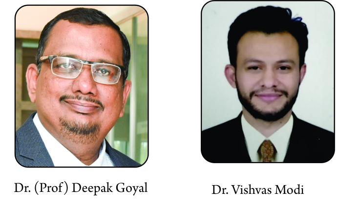
Deepak Goyal, Vishvas Modi

Volume 4 | Issue 1 | Jan – April 2019 | Page 27- 33


Author: Deepak Goyal[1], Vishvas Modi[1]

Saumya Arthroscopy & Sports Knee Clinic,
Ahmedabad, India.

Address of Correspondence
Dr (Prof) Deepak Goyal
Saumya Arthroscopy & Sports Knee Clinic,
Ahmedabad, India.
Email: deepak@knee.in


Abstract

Autologous chondrocyte implantation (ACI) is one such technique that has the ability to provide a hyaline (like) repair of the localized cartilage lesions, even when they are of a big size. However, the procedure must be chosen very wisely because of its stringent indications and contraindications. Decision to do ACI procedure is very crucial and the surgeon must come to the decision after a detailed clinic-radiological examination. Gel based ACI is one such technique that allows a 3-dimensional distribution of the autologous cultured chondrocytes in a scaffold that is made of fibrin glue. The technique takes away the common complications that were associated with 1st and 2nd generation ACI; like graft hypertrophy, poor access to the lesion, membrane suturing, monolayer distribution etc. The purpose of this paper is to discuss the indications, contraindications, decision making and preoperative planning for the gel based autologous chondrocyte implantation technique in detail along with the surgical procedure, postoperative rehabilitation and the possible complications.


References

1. Ossendorf C, Kaps C, Kreuz PC, Burmester GR, Sittinger M, Erggelet C. Treatment of posttraumatic and focal osteoarthritic cartilage defects of the knee with autologous polymer-based three-dimensional chondrocyte grafts: 2-year clinical results. Arthritis Res Ther. 2007;9(2):R41.
2. Oussedik S, Tsitskaris K, Parker D. Treatment of articular cartilage lesions of the knee by microfracture or autologous chondrocyte implantation: a systematic review. Arthroscopy. 2015;31(4):732-744.
3. Goyal D, Keyhani S, Lee EH, Hui JHP. Evidence-Based Status of Microfracture Technique: A Systematic Review of Level I and II Studies. Arthroscopy. 2013;29(9):1579-1588.
4. Bark S, Riepenhof H, Gille J. AMIC Cartilage Repair in a Professional Soccer Player. Case Rep Orthop. 2012;2012:364342.
5. Gille J, Behrens P, Schulz AP, Oheim R, Kienast B. Matrix-Associated Autologous Chondrocyte Implantation: A Clinical Follow-Up at 15 Years. Cartilage. 2016;7(4):309-315.
6. Minas T, Ogura T, Bryant T. Autologous Chondrocyte Implantation. JBJS Essent Surg Tech. 2016;6(2):e24.
7. Bartlett W, Skinner JA, Gooding CR, et al. Autologous chondrocyte implantation versus matrix-induced autologous chondrocyte implantation for osteochondral defects of the knee: a prospective, randomised study. J Bone Joint Surg Br. 2005;87(5):640-645.
8. Behrens P, Bitter T, Kurz B, Russlies M. Matrix-associated autologous chondrocyte transplantation/implantation (MACT/MACI)–5-year follow-up. Knee. 2006;13(3):194-202.
9. Pavesio A, Abatangelo G, Borrione A, et al. Hyaluronan-based scaffolds (Hyalograft C) in the treatment of knee cartilage defects: preliminary clinical findings. Novartis Found Symp. 2003;249:203-217; discussion 229-233, 234-238, 239-241.
10. Schneider U, Rackwitz L, Andereya S, et al. A prospective multicenter study on the outcome of type I collagen hydrogel-based autologous chondrocyte implantation (CaReS) for the repair of articular cartilage defects in the knee. Am J Sports Med. 2011;39(12):2558-2565.
11. Niemeyer P, Albrecht D, Andereya S, et al. Autologous chondrocyte implantation (ACI) for cartilage defects of the knee: A guideline by the working group “Clinical Tissue Regeneration” of the German Society of Orthopaedics and Trauma (DGOU). Knee. 2016;23(3):426-435.
12. Goyal D, Goyal A, Adachi N. Subchondral Bone: Healthy Soil for the Healthy Cartilage. In: Gobbi A, Espregueira-Mendes J, Lane JG, Karahan M, eds. Bio-Orthopaedics. Berlin, Heidelberg: Springer Berlin Heidelberg; 2017:479-486.
13. Cole BJ, Corpus KT, Bajaj S, et al. Prospective Evaluation of Autologous Chondrocyte Implantation Procedure: Minimum Seven-Year Follow-Up (SS-26). Arthroscopy: The Journal of Arthroscopic & Related Surgery. 2011;27(5):e43-e44.
14. Goyal D, Goyal A, Keyhani S, Lee EH, Hui JHP. Evidence-based status of second- and third-generation autologous chondrocyte implantation over first generation: a systematic review of level I and II studies. Arthroscopy. 2013;29(11):1872-1878.
15. Goyal D, Keyhani S, Goyal A, Lee EH, Hui JHP, Vaziri AS. Evidence-Based Status of Osteochondral Cylinder Transfer Techniques: A Systematic Review of Level I and II Studies. Arthroscopy. 2014;30(4):497-505.
16. Pietschmann MF, Horng A, Niethammer T, et al. Cell quality affects clinical outcome after MACI procedure for cartilage injury of the knee. Knee Surg Sports Traumatol Arthrosc. 2009;17(11):1305-1311.
17. Vanlauwe J, Saris DBF, Victor J, et al. Five-year outcome of characterized chondrocyte implantation versus microfracture for symptomatic cartilage defects of the knee: early treatment matters. Am J Sports Med. 2011;39(12):2566-2574.
18. Gillogly SD, Myers TH, Reinold MM. Treatment of full-thickness chondral defects in the knee with autologous chondrocyte implantation. J Orthop Sports Phys Ther. 2006;36(10):751-764.
19. Hamby TS, Gillogly SD, Peterson L. Treatment of patellofemoralarticular cartilage injuries with autologous chondrocyte implantation. Operative Techniques in Sports Medicine. 2002;10(3):129-135.
20. Rosenberg TD, Paulos LE, Parker RD, Coward DB, Scott SM. The forty-five-degree posteroanterior flexion weight-bearing radiograph of the knee. J Bone Joint Surg Am. 1988;70(10):1479-1483.
21. Zbojniewicz AM, Laor T. Imaging of osteochondritis dissecans. Clin Sports Med. 2014;33(2):221-250.
22. Karim AR, Cherian JJ, Jauregui JJ, Pierce T, Mont MA. Osteonecrosis of the knee: review. Ann Transl Med. 2015;3(1):6.
23. Houpt JB, Pritzker KP, Alpert B, Greyson ND, Gross AE. Natural history of spontaneous osteonecrosis of the knee (SONK): a review. Semin Arthritis Rheum. 1983;13(2):212-227.
24. Kellgren JH, Lawrence JS. Radiological assessment of osteo-arthrosis. Ann Rheum Dis. 1957;16(4):494-502.
25. Altman R, Asch E, Bloch D, et al. Development of criteria for the classification and reporting of osteoarthritis. Classification of osteoarthritis of the knee. Diagnostic and Therapeutic Criteria Committee of the American Rheumatism Association. Arthritis Rheum. 1986;29(8):1039-1049.
26. Spector TD, Hart DJ, Byrne J, Harris PA, Dacre JE, Doyle DV. Definition of osteoarthritis of the knee for epidemiological studies. Ann Rheum Dis. 1993;52(11):790-794.
27. Goyal D, Goyal A, Adachi N. Joint Preservation Surgery for Medial Compartment Osteoarthritis. Arthrosc Tech. 2017;6(3):e717-e728.
28. Recht MP, Resnick D. Magnetic resonance imaging of articular cartilage: an overview. Top Magn Reson Imaging. 1998;9(6):328-336.
29. De Smet AA, Fisher DR, Graf BK, Lange RH. Osteochondritis dissecans of the knee: value of MR imaging in determining lesion stability and the presence of articular cartilage defects. AJR Am J Roentgenol. 1990;155(3):549-553.
30. Lecouvet FE, van de Berg BC, Maldague BE, et al. Early irreversible osteonecrosis versus transient lesions of the femoral condyles: prognostic value of subchondral bone and marrow changes on MR imaging. AJR Am J Roentgenol. 1998;170(1):71-77.
31. Mont MA, Baumgarten KM, Rifai A, Bluemke DA, Jones LC, Hungerford DS. Atraumatic osteonecrosis of the knee. J Bone Joint Surg Am. 2000;82(9):1279-1290.
32. Björkengren AG, AlRowaih A, Lindstrand A, Wingstrand H, Thorngren KG, Pettersson H. Spontaneous osteonecrosis of the knee: value of MR imaging in determining prognosis. AJR Am J Roentgenol. 1990;154(2):331-336.
33. Healy WL. Osteonecrosis of the knee detected only by magnetic resonance imaging. Orthopedics. 1991;14(6):703-704.
34. Goyal D, Goyal A, Brittberg M. Consideration of religious sentiments while selecting a biological product for knee arthroscopy. Knee Surg Sports Traumatol Arthrosc. 2013;21(7):1577-1586.
35. Goyal D, Palkhiwala B. Cover Image. Arthroscopy: The Journal of Arthroscopic & Related Surgery. 2013;29(3):A9.
36. Goyal D. The overlay ACI technique for extra-large osteochondral lesions of the knee joint. 2nd Congress of Asian Cartilage Repair Society at Seoul, Korea, 2014.
37. Choi N-Y, Kim B-W, Yeo W-J, et al. Gel-type autologous chondrocyte (Chondron) implantation for treatment of articular cartilage defects of the knee. BMC Musculoskelet Disord. 2010;11:103.
38. Gillogly SD, Voight M, Blackburn T. Treatment of articular cartilage defects of the knee with autologous chondrocyte implantation. J Orthop Sports Phys Ther. 1998;28(4):241-251.
39. Reinold MM, Wilk KE, Dugas JR, Cain EL, Gillogly SD. Rehabilitation Guidelines: Autologous Chondrocyte Implantation Using Carticel. Cambridge, MA: Genzyme Biosurgery; 2004.
40. Reinold MM, Wilk KE, Macrina LC, Dugas JR, Cain EL. Current concepts in the rehabilitation following articular cartilage repair procedures in the knee. J Orthop Sports Phys Ther. 2006;36(10):774-794.
41. Peterson L, Minas T, Brittberg M, Nilsson A, Sjögren-Jansson E, Lindahl A. Two- to 9-year outcome after autologous chondrocyte transplantation of the knee. Clin Orthop Relat Res. 2000;(374):212-234.
42. Peterson L, Brittberg M, Kiviranta I, Akerlund EL, Lindahl A. Autologous chondrocyte transplantation. Biomechanics and long-term durability. Am J Sports Med. 2002;30(1):2-12.
43. Peterson L, Vasiliadis HS, Brittberg M, Lindahl A. Autologous chondrocyte implantation: a long-term follow-up. Am J Sports Med. 2010;38(6):1117-1124.
44. Pareek A, Carey JL, Reardon PJ, Peterson L, Stuart MJ, Krych AJ. Long-Term Outcomes after Autologous Chondrocyte Implantation: A Systematic Review at Mean Follow-Up of 11.4 Years. Cartilage. 2016;7(4):298-308.
45. Kim MK, Choi SW, Kim SR, Oh IS, Won MH. Autologous chondrocyte implantation in the knee using fibrin. Knee Surg Sports Traumatol Arthrosc. 2010;18(4):528-534.
46. Wood JJ, Malek MA, Frassica FJ, et al. Autologous cultured chondrocytes: adverse events reported to the United States Food and Drug Administration. J Bone Joint Surg Am. 2006;88(3):503-507.
47. Niemeyer P, Pestka JM, Kreuz PC, et al. Characteristic complications after autologous chondrocyte implantation for cartilage defects of the knee joint. Am J Sports Med. 2008;36(11):2091-2099.


How to Cite this article: Goyal & Modi. Gel Based Autologous Chondrocyte Implantation: The Surgical Technique. Asian Journal Arthroscopy. Jan-April 2019;4(1):27-33 .


(Abstract)      (Full Text HTML)      (Download PDF)


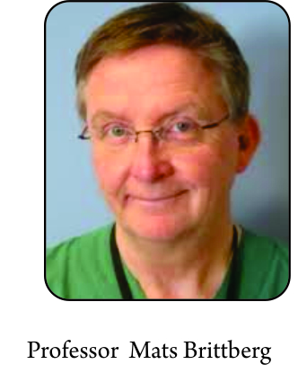
Professor Mats Brittberg

Volume 4 | Issue 1 | Jan – April 2019 | Page 23- 26


Author: Professor Mats Brittberg[1]

[1] Cartilage Research Unit, University of Gothenburg, Region Halland Orthopaedics, Kungsbacka Hospital, S-434 80 Kungsbacka Sweden.

Address of Correspondence
Professor- Mats Brittberg
Cartilage Research Unit, University of Gothenburg, Region Halland Orthopaedics, Kungsbacka Hospital, S-434 80 Kungsbacka Sweden.
Email: Mats.brittberg@telia.com


Abstract

Autologous chondrocyte implantation (ACI) has been used clinically since 1987. Most reports on ACI are on the first and second generation ACI with cells in suspension under a sutured membrane and performed with open surgery. Today’s 3rd and 4th generation ACI with cells seeded and grown in or on scaffolds prior to implantation opens up for transarthroscopic implantations. Transarthroscopic surgery reduces the morbidity for the patients and also fastens up the rehab process. In this paper, techniques used for scaffold-based ACI are presented.


References

1.Brittberg M, Lindahl A, Nilsson A et al.Treatment of deep cartilage defects in the knee with autologous chondrocyte transplantation. New England Journal of Medicine 1994; 331:889-895.
2.Brittberg M.Cartilage repair. On cartilaginous tissue engineering with the emphasis on chondrocyte transplantation. Thesis. University of Gothenburg.1996.
3.Brittberg M. Cell carriers as the next generation of cell therapy for cartilage repair: a review of the matrix-induced autologous chondrocyte implantation procedure.Am J Sports Med. 2010; Jun;38(6):1259-71.
4.Steinwachs M. New technique for cell-seeded collagen-matrix-supported autologous chondrocyte transplantation. Arthroscopy. 2009; Feb;25(2):208-11.
5.Hendriks JA, Moroni L, Riesle et al. The effect of scaffold-cell entrapment capacity and physico-chemical properties on cartilage regeneration. Biomaterials. 2013 Jun;34(17):4259-65
6.de Windt TS, Vonk LA, Slaper-Cortenbach ICM, Nizak R, van Rijen MHP, Saris DBF. Allogeneic MSCs and Recycled Autologous Chondrons Mixed in a One-Stage Cartilage Cell Transplantion: A First-in-Man Trial in 35 PatientsStem Cells. 2017 Aug;35(8):1984-1993
7.Brittberg M. Cartilage fragment implantation. . In Cartilage Surgery. An operative manual. Eds Brittberg M, Gersoff W. Elsevier Saunders Philadelphia,2011; 181-191.
8.Brittberg M, Concaro S. Transarthroscopic implantation of Hyalograft (Hyaff 11) with autologous chondrocytes. In Cartilage Surgery. An operative manual. Eds Brittberg M, Gersoff W. Elsevier Saunders Philadelphia,2011; 147-160.
9. Nehrer S, Dorotka R, Domayer S et al. Treatment of full-thickness chondral defects with hyalograft C in the knee: a prospective clinical case series with 2 to 7 years’ follow-up. Am J Sports Med. 2009; Nov;37 Suppl 1:81S-87S
10.Vascellari A, Rebuzzi E, Schiavetti et al. Implantation of matrix-induced autologous chondrocyte (MACI(®)) grafts using carbon dioxide insufflation arthroscopy. Knee Surg Sports Traumatol Arthrosc. 2014 Jan;22(1):219-25.
11.Marcacci M, Berruto M, Brocchetta D et al. Articular cartilage engineering with Hyalograft C: 3-year clinical results. Clin Orthop Relat Res.2005; Jun;(435):96-105
12.Della Villa S, Kon E, Filardo G et al. Does intensive rehabilitation permit early return to sport without compromising the clinical outcome after arthroscopic autologous chondrocyte implantation in highly competitive athletes? Am J Sports Med. 2010; Jan;38(1):68-77.


How to Cite this article: Brittberg M. Scaffold based autologous chondrocyte implantation: The surgical technique. Asian Journal Arthroscopy. Jan-April 2019;4(1):23-26 .


(Abstract)      (Full Text HTML)      (Download PDF)


Rajkumar Amaravathi, Renato Andrade, Ricardo Bastos, João Espregueira-Mendes

Volume 4 | Issue 1 | Jan – April 2019 | Page 15-22


Author: Rajkumar Amaravathi [1], Renato Andrade [2,3,4], Ricardo Bastos [2,3,5,6], João Espregueira-Mendes [2]

[1] Department Of Orthopedics, Division of Arthroscopy and Sports Surgery, St. John’s Medical College and Hospital, Bangalore 560034,India
[2] Clínica do Dragão, Espregueira-Mendes Sports Centre – FIFA Medical Centre of Excellence, Porto, Portugal.
[3] Dom Henrique Research Centre, Porto, Portugal.
[4] Faculty of Sports, University of Porto, Porto, Portugal.
[5] Fluminense Federal University, Niteroi, Brazil.
[6] ICVS/3B’s-PT Government Associate Laboratory, Guimarães, Portugal.
[7] School of Medicine, Minho University, Braga, Portugal.

Address of Correspondence
Dr João Espregueira-Mendes,
M.D., Ph.D.; Clínica do Dragão, Espregueira-Mendes Sports Centre – FIFA Medical Centre of Excellence,
Estádio do Dragão, Entrada Nascente, Piso -3, 4350-415, Porto, Portugal.
Email: espregueira@dhresearchcentre.com


Abstract

Osteochondral autologous transplantation is a surgical procedure that involves the transplant of the autologous cartilage from the non-weight bearing areas of the knee to the articular defect. It has the advantage of being a single stage procedure, repairs the subchondral bone, provides hyaline cartilage and allows a fast return to play. It is indicated for small and medium-sized defects, but the mosaicplasty technique allows treating defects up to 9 cm2. A major disadvantage of this technique is the donor site morbidity associated with the graft harvesting. To overcome this drawback, we harvest the autografts from the upper tibio-fibular joint with low or none donor site morbidity. Osteochondral autologous transplantation and mosaicplasty procedures remain an excellent option for small to medium osteochondral injuries resulting in long-term good to excellent clinical and imaging outcomes.Osteochondral autologous transplantation is a surgical procedure that involves the transplant of the autologous cartilage from the non-weight bearing areas of the knee to the articular defect. It has the advantage of being a single stage procedure, repairs the subchondral bone, provides hyaline cartilage and allows a fast return to play. It is indicated for small and medium-sized defects, but the mosaicplasty technique allows treating defects up to 9 cm2. A major disadvantage of this technique is the donor site morbidity associated with graft harvesting. To overcome this drawback, we harvest the autografts from the upper tibio-fibular joint with low or none donor site morbidity. Osteochondral autologous transplantation and mosaicplasty procedures remain an excellent option for small to medium osteochondral injuries resulting in long-term good to excellent clinical and imaging outcomes.


References

1. Curl WW, Krome J, Gordon ES, Rushing J, Smith BP, Poehling GG. Cartilage injuries: a review of 31,516 knee arthroscopies. Arthroscopy. 1997;13:456-460.
2. Cognault J, Seurat O, Chaussard C, Ionescu S, Saragaglia D. Return to sports after autogenous osteochondral mosaicplasty of the femoral condyles: 25 cases at a mean follow-up of 9 years. Orthop Traumatol Surg Res. 2015;101:313-317.
3. Heijink A, Gomoll AH, Madry H, Drobnič M, Filardo G, Espregueira-Mendes J, Van Dijk CN. Biomechanical considerations in the pathogenesis of osteoarthritis of the knee. Knee Surg Sports Traumatol Arthrosc. 2012;20:423-435.
4. Gomoll A, Filardo G, De Girolamo L, Esprequeira-Mendes J, Marcacci M, Rodkey W, Steadman R, Zaffagnini S, Kon E. Surgical treatment for early osteoarthritis. Part I: cartilage repair procedures. Knee Surg Sports Traumatol Arthrosc. 2012;20:450-466.
5. Vannini F, Spalding T, Andriolo L et al. Sport, and early osteoarthritis: the role of sport in aetiology, progression, and treatment of knee osteoarthritis. Knee Surg Sports Traumatol Arthrosc. 2016;24:1786-1796.
6. Heir S, Nerhus TK, Røtterud JH, Løken S, Ekeland A, Engebretsen L, Årøen A. Focal Cartilage Defects in the Knee Impair Quality of Life as Much as Severe Osteoarthritis: A Comparison of Knee Injury and Osteoarthritis Outcome Score in 4 Patient Categories Scheduled for Knee Surgery. Am J Sports Med. 2009;38:231-237.
7. Krych AJ, Gobbi A, Lattermann C, Nakamura N. Articular cartilage solutions for the knee: present challenges and future direction. JISAKOS. 2016;1:93-104.
8. Kreuz PC, Steinwachs MR, Erggelet C, Krause SJ, Konrad G, Uhl M, Südkamp N. Results after microfracture of full-thickness chondral defects in different compartments in the knee. Osteoarthritis Cartilage. 2006;14:1119-1125.
9. Gudas R, Kalesinskas RJ, Kimtys V, Stankevic̆ius E, Tolius̆is V, Bernotavic̆ius G, Smailys A. A prospective randomized clinical study of mosaic osteochondral autologous transplantation versus microfracture for the treatment of osteochondral defects in the knee joint in young athletes. Arthroscopy. 2005;21:1066-1075.
10. Peterson L, Minas T, Brittberg M, Nilsson A, Sjögren-Jansson E, Lindahl A. Two- to 9-Year Outcome After Autologous Chondrocyte Transplantation of the Knee. Clin Orthop Relat Res. 2000;374:212-234.
11. Hangody L, Fuels P. Autologous osteochondral mosaicplasty for the treatment of full-thickness defects of weight-bearing joints: ten years of experimental and clinical experience. J Bone Joint Surg Am. 2003;85-A Suppl 2:25-32.
12. Andrade R, Vasta S, Pereira R, Pereira H, Papalia R, Karahan M, Oliveira JM, Reis RL, Espregueira-Mendes J. Knee donor-site morbidity after mosaicplasty–a systematic review. J Exp Orthop. 2016;3:31.
13. Espregueira-Mendes J, Pereira H, Sevivas N, Varanda P, Da Silva MV, Monteiro A, Oliveira JM, Reis RL. Osteochondral transplantation using autografts from the upper tibio-fibular joint for the treatment of knee cartilage lesions. Knee Surg Sports Traumatol Arthrosc. 2012;20:1136-1142.
14. Hangody L, Vasarhelyi G, Hangody LR, Sukosd Z, Tibay G, Bartha L, Bodo G. Autologous osteochondral grafting-technique and long-term results. Injury. 2008;39 Suppl 1:S32-39.
15. Camp CL, Stuart MJ, Krych AJ. Current concepts of articular cartilage restoration techniques in the knee. Sports Health. 2014;6:265-273.
16. Brittberg M, Winalski CS. Evaluation of cartilage injuries and repair. J Bone Joint Surg Am. 2003;85-A Suppl 2:58-69.
17. Trattnig S, Domayer S, Welsch GW, Mosher T, Eckstein F. MR imaging of cartilage and its repair in the knee-a review. Eur Radiol. 2009;19:1582-1594.
18. Messner K, Maletius W. The long-term prognosis for severe damage to weight-bearing cartilage in the knee: a 14-year clinical and radiographic follow-up in 28 young athletes. Acta Orthop Scand. 1996;67:165-168.
19. Garretson RB, Katolik LI, Verma N, Beck PR, Bach BR, Cole BJ. Contact Pressure at Osteochondral Donor Sites in the Patellofemoral Joint. Am J Sports Med. 2004;32:967-974.
20. Ahmad CS, Cohen ZA, Levine WN, Ateshian GA, Van CM.Biomechanical and Topographic Considerations for Autologous Osteochondral Grafting in the Knee. Am J Sports Med; 2001;29:201-206.
21. Thaunat M, Couchon S, Lunn J, Charrois O, Fallet L, Beaufils P. Cartilage thickness matching of selected donor and recipient sites for osteochondral autografting of the medial femoral condyle. Knee Surg Sports Traumatol Arthrosc. 2007;15:381-386.
22. Keeling JJ, Gwinn DE, McGuigan FX. A comparison of open versus arthroscopic harvesting of osteochondral autografts. Knee. 2009;16:458-462.
23. Duchow J, Hess T, Kohn D. Primary stability of press-fit-implanted osteochondral grafts: influence of graft size, repeated insertion, and harvesting technique. Am J Sports Med. 2000;28:24-27.
24. Makino T, Fujioka H, Terukina M, Yoshiya S, Matsui N, Kurosaka M. The effect of graft sizing on osteochondral transplantation. Arthroscopy. 2004;20:837-840.
25. Huang FS, Simonian PT, Norman AG, Clark JM. Effects of small incongruities in a sheep model of osteochondral autografting. Am J Sports Med. 2004;32:1842-1848.
26. Patil S, Butcher W, D’lima DD, Steklov N, Bugbee WD, Hoenecke HR. Effect of osteochondral graft insertion forces on chondrocyte viability. Am J Sports Med. 2008;36:1726-1732.
27. Kock N, van Susante J, Wymenga A, Buma P. Histological evaluation of a mosaicplasty of the femoral condyle—retrieval specimens obtained after total knee arthroplasty—a case report. Acta Orthop Scand. 2004;75:505-508.
28. Ahmad CS, Guiney WB, Drinkwater CJ. Evaluation of donor site intrinsic healing response in autologous osteochondral grafting of the knee. Arthroscopy. 2002;18:95-98.
29. Espregueira-Mendes J, Andrade R, Monteiro A, Pereira H, da Silva MV, Oliveira JM, Reis RL. Mosaicplasty Using Grafts From the Upper Tibiofibular Joint. Arthrosc Tech. 2017;6:e1979-e1987.
30. Robert H, Lambotte J, Flicoteaux R. Arthroscopic measurements of cartilage lesions of the knee condyle. Principles and experimental validation of a new method. Cartilage. 2011;2: 237–245
31. Bobić V.Arthroscopic osteochondral autograft transplantation in anterior cruciate ligament reconstruction: a preliminary clinical study. Knee Surg Sports Traumatol Arthrosc. 1996;3:262-264.
32. Evans PJ, Miniaci A, Hurtig MB. Manual punch versus power harvesting of osteochondral grafts. Arthroscopy. 2004;20:306-310.
33. Hurtig M, Evans P, Pearce S, Clarnette R, Miniaci A The effect of graft size and number on the outcome of mosaic arthroplasty resurfacing: an experimental model in sheep. In: Transactions, 18th Annual Meeting of the Arthroscopy Association of North America, Vancouver, 1999:16-17.
34. Bader S, Miniaci A. Mosaicplasty. Orthopedics. 2011;32:678-678.
35. Espregueira-Mendes J, Da Silva MV. Anatomy of the proximal tibiofibular joint. Knee Surg Sports Traumatol Arthrosc. 2006;14:241-249.
36. Andrade R, Pereira R, Bastos R, Saavedra C, Pereira H, Laver L, Landreau P, Espregueira-Mendes J. Management of Cartilage Injuries in Handball. In: Laver, L., Landreau, P., Seil, R., Popovic, N. (Eds). Handball Sports Medicine: Basic Science, Injury Management and Return to Sport. Springer; 2018, p. 325-340.
37. Andrade R, Pereira R, Bastos R, Pereira H, Oliveira JM, Reis RL, Espregueira-Mendes J. Return to Play Following Cartilage Injuries. In: Musahl, V., Karlsson, J., Krutsch, W., Mandelbaum, B.R., Espregueira-Mendes, J., d’Hooghe, P. (Eds). Return to Play in Football: An Evidence-based Approach. Springer; 2018, p. 593-610.
38. Mithoefer K, Hambly K, Logerstedt D, Ricci M, Silvers H, Villa SD. Current concepts for rehabilitation and return to sport after knee articular cartilage repair in the athlete. J Orthop Sports Phys Ther. 2012; 42:254-273.
39. Horas U, Pelinkovic D, Herr G, Aigner T, Schnettler R. Autologous chondrocyte implantation and osteochondral cylinder transplantation in cartilage repair of the knee joint: a prospective, comparative trial. J Bone Joint Surg Am. 2013;85:185-192.
40. Bentley G, Biant L, Carrington R, Akmal M, Goldberg A, Williams A, Skinner J, Pringle J. A prospective, randomised comparison of autologous chondrocyte implantation versus mosaicplasty for osteochondral defects in the knee. J Bone Joint Surg Br. 2003;85:223-230.
41. Jungmann PM, Gersing AS, Baumann F et al. Cartilage repair surgery prevents progression of knee degeneration. Knee Surg Sports Traumatol Arthrosc. 2018.
42. Solheim E, Hegna J, Strand T, Harlem T, Inderhaug E. Randomized study of long-term (15-17 years) outcome after microfracture versus mosaicplasty in knee articular cartilage defects. Am J Spots Med. 2018;46:826-831.
43. Gudas R, Gudaite A, Mickevicius T, Masiulis N, Simonaityte R, Cekanauskas E, Skurvydas A. Comparison of osteochondral autologous transplantation, microfracture, or debridement techniques in articular cartilage lesions associated with anterior cruciate ligament injury: a prospective study with a 3-year follow-up. Arthroscopy. 2013;29:89-97.
44. Gudas R, Gudaite A, Pocius A, Gudiene A, Cekanauskas E, Monastyreckiene E, Basevicius A. Ten-year follow-up of a prospective, randomized clinical study of mosaic osteochondral autologous transplantation versus microfracture for the treatment of osteochondral defects in the knee joint of athletes. Am J Sports Med. 2012;40:2499-2508.
45. Gudas R, Simonaityte R, Cekanauskas E, Tamosiunas R. A prospective, randomized clinical study of osteochondral autologous transplantation versus microfracture for the treatment of osteochondritis dissecans in the knee joint in children. J Pediatr Orthop. 2012;29:741-748.
46. Lim HC, Bae JH, Song SH, Park YE, Kim SJ. Current treatments of isolated articular cartilage lesions of the knee achieve similar outcomes. Clin Orthop Relat Res. 2012;470:2261-2267.
47. Ulstein S, Aroen A, Rotterud JH, Loken S, Engebretsen L, Heir S. Microfracture technique versus osteochondral autologous transplantation mosaicplasty in patients with articular chondral lesions of the knee: a prospective randomized trial with long-term follow-up. Knee Surg Sports Traumatol Arthrosc. 2014;22:1207-1215.
48. Mithoefer K, Hambly K, Della Villa S, Silvers H, Mandelbaum BR. Return to sports participation after articular cartilage repair in the knee: scientific evidence. Am J Sports Med. 2009;37 Suppl 1:167s-176s.
49. Andrade R, Vasta S, Papalia R, Pereira H, Oliveira JM, Reis RL, Espregueira-Mendes J. Prevalence of articular cartilage lesions and surgical clinical outcomes in football (soccer) players’ knees: a systematic review. Arthroscopy. 2016;32:1466-1477.
50. Krych AJ, Pareek A, King AH, Johnson NR, Stuart MJ, Williams RJ, 3rd. Return to sport after the surgical management of articular cartilage lesions in the knee: a meta-analysis. Knee Surg Sports Traumatol Arthrosc. 2017;25:3186-3196.


How to Cite this article: Rajkumar Amaravathi R, Andrade R, Bastos R, Espregueira-Mendes J. The Mosaicplasty / OAT procedure: Technique, Pearls and Pitfalls. Asian Journal Arthroscopy. Jan-April 2019;4(1):15-22 .


(Abstract)      (Full Text HTML)      (Download PDF)


Endoscopic Plantar Fasciotomy with Gastrocnemius Recession for Chronic Plantar Fasciitis

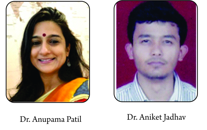
Anupama Patil, Aniket Jadhav

Volume 4 | Issue 1 | Jan – April 2019 | Page 4-8


Author: Anupama Patil[1], Aniket Jadhav[1]

STAR imaging and research centre, Joshi Hospital Campus Opposite Kamla Nehru Park, Erandwane, Pune, Maharashtra.

Address of Correspondence
Dr Anupama Patil
STAR imaging and research centre, Joshi Hospital Campus Opposite Kamla Nehru Park, Erandwane, Pune, Maharashtra 411004
Email: anupama.patil2003@gmail.com


Abstract

Chondral injuries can occur in an isolated manner or, more commonly, in association with osseous or soft tissue injuries. Accurate pre-knowledge of the chondral injury and associated injuries help the orthopedic surgeon in planning appropriate treatment procedures. Advances in various treatment techniques for chondral defects places paramount importance on the identification, and quantification of these injuries. Through this article, we present a review of literature regarding Magnetic resonance imaging assessment of chondral injuries, also addressing the scan parameters used, advances in imaging for cartilage, role of Magnetic resonance imaging in postoperative follow-up, comparison of accuracy of Magnetic resonance imaging with arthroscopy as well as the roles of ultrasonography and computed tomography in evaluation of articular cartilage.
Magnetic resonance imaging has an indispensable role in the pre-arthroscopic work-up and post-arthroscopic follow-up of chondral injuries. It gives an accurate knowledge of chondral defects/ injuries, staging of lesions, evaluating subchondral bone, assessing adjacent cartilage, identifying loose bodies in remote recesses likely to be missed on arthroscopy, and identifying another ligament/meniscal tears. It is also useful in assessing the donor and recipient sites in
The post-arthroscopic workup following cartilage repair. Ultrasound arthroscopy is a new quantitative intra-operative imaging modality, still not widely used. Computed tomography doesn’t image the cartilage directly but plays an important ancillary role in the evaluation of subchondral bone and identification of location and size of loose bodies.


References

1. Michel D. Crema, Frank W. Roemer et al.Articular cartilage in the knee: current MR imaging techniques and applications in clinical practice and research.Radiographics.2011;31(1):37-62.
2. Tallal C. Mamisch, Siegfried Trattnig et al. Musculoskeletal Imaging : Quantitative T2 Mapping of Knee Cartilage Differentiation of Healthy Control Cartilage and Cartilage Repair Tissue in the Knee with Unloading—Initial Results.Radiology.2010;254(3):818-26.
3. Garry E. Gold, Christina A. Chen et al. Recent Advances in MRI of Articular Cartilage.American Journal of Roentgenology.2009;193:628-38 .
4. Yon Sun Choi, Hollis G. Potter, Tong Jin Chun. MR imaging of Cartilage Repair in the Knee and Ankle. Radiographics.2008;28(4):1043-59.
5. Christiaan JA van Bergen, Rogier Gerards et al. Diagnosing, planning and evaluating osteochondral ankle defects with imaging modalities.World J Orthop.2015;6(11):944–53.
6. Pekko Penttilä, Jukka Liukkonen et al.Diagnosis of Knee Osteochondral Lesions With Ultrasound Imaging.Arthrosc Tech.2015;4(5):e429–e433.
7. Kira D. Novakofski, Sarah L. Pownder et al. High-Resolution Methods for Diagnosing Cartilage Damage In Vivo. Cartilage. 2016; 7(1): 39–51.
8. Bhawan K Paunipagar, DD Rasalkar et al.Imaging of articular cartilage.Indian J Radiol Imaging.2014;24(3):237-48.
9. Irmak Durur-Subasi, Afak Durur-Karakaya et al.Osteochondral Lesions of Major Joints.Eurasian J Med.2015;47:138-44.
10. Marcelo Bordalo Rodrigues, Gilberto Luís Camanho.MRI EVALUATION OF KNEE CARTILAGE. Rev Bras Ortop.2010;45(4):340-6.
11. Candace L. White, Nancy A. Chauvin et al.MRI of Native Knee Cartilage Delamination Injuries.American Journal of Roentgenology.2017;209(5):W317-W321.
12. Richard Kijowski.Clinical Cartilage Imaging of the Knee and Hip Joints.Americal Journal of Roentgenology.2010;195:618-28.
13. Richard Kijowski, Donna G. Blankenbaker et al. Comparison of 1.5- and 3.0-T MR Imaging for Evaluating the Articular Cartilage of the Knee Joint.Radiology.2009;250(3):839-848.


How to Cite this article: Patil A, Jadhav A. Imaging for Cartilage injuries. Asian Journal Arthroscopy. Jan-April 2019;4(1):4-8 .


(Abstract)      (Full Text HTML)      (Download PDF)


Endoscopic Plantar Fasciotomy with Gastrocnemius Recession for Chronic Plantar Fasciitis

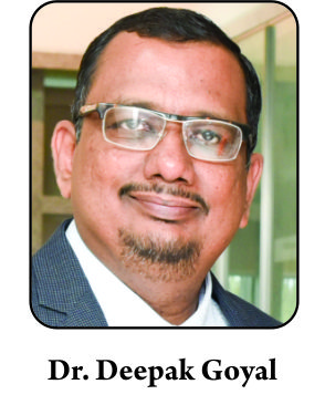
Dr. Deepak Goyal

Volume 4 | Issue 1 | Jan – April 2019 | Page 2-3


Author: Deepak Goyal [1]

Consultant Cartilage and Sports Knee Surgeon, Saumya Arthroscopy & Sports Knee Clinic, Ahmedabad, India.

Address of Correspondence
Dr. Deepak Goyal,
Consultant Cartilage and Sports Knee Surgeon
Saumya Arthroscopy & Sports Knee Clinic,
Ahmedabad, India.
Email: deepak@knee.in


Abstract

Hunter [1] in AD 1742 had observed, ‘Cartilage once damaged cannot heal’ and the orthopedic community continued to believe in his statement for over 200 years. There were some scattered attempts and observations to heal the cartilage in the later-half of the twentieth century. We have seen an enthusiastic interest with many new modalities showing varying degrees of cartilage repair success in the last 25 years. While traveling across Asian countries, I come across many surgeons who have an active interest in the cartilage repair. Young enthusiast surgeons are keen to learn techniques and develop skills for various cartilage repair procedures. But, many of them are unable to do more than the microfracture technique, the first line of treatment [2]. Asian Journal of Arthroscopy, is keen to infuse the basic know-how of commonly done procedures along with a stress on practical approach towards the procedures. Literature is abundant about each of the cartilage repair procedures, but a ‘beginner cartilage surgeon’ would like to know the basics of case selection, decision making, surgical techniques, and tips, along with the possible complications. This special issue on cartilage repair invited different surgeons/ doctors who are doing pioneer work in their respective fields of cartilage repair.

Patil and Jadhav [3] have started this edition of AJA with a detailed understanding of various sequences of MRI that are important for the diagnosis of the cartilage lesions. They have not only described the various zones of cartilage on MRI but have also discussed in detail about the T2 mapping. The various MRI characteristics of damaged cartilage and repaired cartilage are also discussed along with the MOCART score.

Devin Leland et at [4] have written a nice article on the microfracture technique that is specifically indicated for the small size lesions. They have given a detailed discussion on the indications, surgical steps, and the site-specific rehabilitation program; while using the microfracture technique. They have also discussed short and long-term results of the microfracture technique along with a brief discussion on various systematic reviews. To conclude, they have also compared the results of the subchondral drilling technique and the osteochondral grafting.

Amaravathi et al [5] advocate osteochondral cylinder transfer technique for mid-size cartilage lesions. They have put a special emphasis on the proper case selection, pre-operative planning, and the surgical technique, along with the detailed tabulated pearls and pitfalls for each. The discussion about long-term results (15 years) of the osteochondral cylinder transfer procedures by various authors is quite promising in favor of the technique. Their suggestion on the use of the proximal tibiofibular joint as the source of the graft will be interesting to watch, with the long-term results in the future.

Mats Brittberg [6] has written a very nice article on 3rd generation ACI and has tried to remove the confusion between the nomenclature of various methods that fall under the ambit of 3rd generation ACI. He has also introduced the 4th generation ACI with this article that will allow the surgeons to use ACI as a single stage surgery.

Goyal and Modi [7] have advocated the use of the gel based ACI procedure, another 3rd generation ACI, mainly for the large chondral lesions with a cautious use in the extra-large lesions. They have heavily emphasised on a very careful selection of the patient using stringent guidelines and a preoperative planning methodology. A detailed step-by-step surgical technique discussion and a postoperative rehabilitation program is also discussed, along with an insight into the possible complications.

Herman et al [8] have stressed the importance of a single stage technique of cartilage repair using hyaluronic acid-based scaffold with the bone marrow aspirate concentrate. They have described the surgical technique along with a detailed understanding of the various phases of the rehabilitation program.

I hope, many youngsters as well as seniors will find this special issue on cartilage repair, useful.


References

1. Hunter W. Of the structure and diseases of articulating cartilages, by William Hunter, surgeon. Philos Trans R Soc Lond 1742;42:514-521.
2. Goyal D, Keyhani S, Lee EH, Hui JHP. Evidence-Based Status of Microfracture Technique: A Systematic Review of Level I and II Studies. Arthroscopy. 2013;29(9):1579-1588.
3. Patil A, Jadhav A. Imaging for Cartilage injuries. Asian Journal Arthroscopy. Jan-April 2019;4(1): 4-8.
4. Leland DP, Bernard CD, Krych AJ. The Microfracture Technique: Pearls and Pitfalls. Asian Journal Arthroscopy. Jan-April 2019;4(1):9-14
5. Amaravathi R, Andrade R, Bastos R, Espregueira-Mendes J. The Mosaicplasty/ OAT procedure: Technique, Pearls, and Pitfalls. Asian Journal Arthroscopy. Jan-April 2019;4(1): 15-22.
6. Brittberg M. Scaffold-based autologous chondrocyte implantation: The surgical technique. Asian Journal Arthroscopy. Jan-April 2019;4(1):23-26
7. Goyal & Modi. Gel-Based Autologous Chondrocyte Implantation: The Surgical Technique. Asian Journal Arthroscopy. Jan-April 2019;4(1):27-33.
8. Herman K, Kumar V, Szwedowski D, Chierici L, Gobbi A. HA-BMAC: The Surgical Technique, Pearls and Pitfalls. Asian Journal Arthroscopy. Jan-April 2019;4(1):34-37


How to Cite this article: Goyal D. A Surgical Perspective to the Modern Cartilage Repair Techniques. Asian Journal of Arthroscopy Jan – April 2019;4(1):2-3


(Abstract)      (Full Text HTML)      (Download PDF)


Endoscopic Plantar Fasciotomy with Gastrocnemius Recession for Chronic Plantar Fasciitis